Johns Hopkins University researchers have uncovered exactly how spiders build webs, using night vision and artificial intelligence to track and record every movement of their eight legs as they work in the dark.
A new understanding has been revealed of how spiders can create webs, and structures of such grace, complexity and geometric precision by these magnificent creatures. The results of the researchers’ study were published in the high-impact (2020 Impact Factor=10.834) journal of Cell Press, a respected publishing house, called Current Biology.
Andrew Gordus, the senior author of the study, said: “I first started this topic while birding with my son. After seeing a magnificent net, I thought, ‘If you went to a zoo and saw a chimpanzee doing this, you would think it was an amazing and impressive chimpanzee.’ This is even more surprising because “A spider’s brain is so small, and I’m frustrated that we don’t know more about how this unusual behaviour occurs. We’ve now described the choreography of web formation that has never been done in such fine resolution for any animal architecture.” this made a statement.
Spiders, which blindly construct webs using only their sense of touch, have fascinated people for centuries. Andrew Gordus explained that the first step to understanding how the relatively small brains of these animal architects support high-level construction projects is to systematically document, analyze and record actions related to behaviours and motor skills that have never been done before.
In their study, the research team used a spider species (Uloborus diversus) native to the western United States that is small enough to fit comfortably on a fingertip. He designed a lab environment with infrared cameras and lights to observe the spiders during their nocturnal web-building work.
With this setup, they tracked and recorded six spiders as they formed their webs each night. They tracked and documented millions of individual leg movements with machine vision software explicitly designed to detect limb movement.
The researchers found that their web-forming behaviour was quite similar among spiders, such that they were able to predict the part of the web that a spider is working on by simply seeing the position of one leg.
“Even though the final structure is slightly different, the rules they use to build the network are the same. They all use the same rules, which confirms that the rules are encoded in their brains. Now we want to know how these rules are encoded at the level of neurons.”
Abel Cover, a lead author of the study, said: “The spider is fascinating because here you have an animal with a brain built on the same basic building blocks as ours, and this work could offer clues to how we might understand larger brain systems, including humans, and I think it’s very exciting. ” made the statement.
Study authors included Nicholas Wilkerson, a former John Hopkins undergraduate and now a graduate student at Atlantic Veterinary College, and Jeremy Miller, a graduate student at Johns Hopkins.
Further Reading: Abel Corver, Nicholas Wilkerson, Jeremiah Miller, Andrew Gordus. Distinct movement patterns generate stages of spider web building. Current Biology, 2021; DOI: 10.1016/j.cub.2021.09.030
Source: Johns Hopkins University. “Spiders’ web secrets unraveled: Researchers document every step of spider-web building.” ScienceDaily. www.sciencedaily.com/releases/2021/11/211101105356.htm (accessed November 9, 2021).
The existence of molecular chirality in the substances in the universe and the stereoisomer properties of molecules with stereogenic centres have been very influential in the development of organic chemistry and related sciences. The reason why chirality is important as biological activity is that molecular symmetry dominates biological events.
Although chirality is not essential for bioactivity, there are great differences in the activities of enantiomers in bioactive molecules with stereogenic centres such as drugs, flavourings, and food additives. The molecular components of living organisms are mostly chiral and these molecules have a dominant role in their interaction with bioactive substances. Receptors or free nerve endings enable all living systems to recognize and perceive all changes in the external environment they live in, such as chemical, physical, electrical, etc. that occur in their internal environment. Regardless of their functions in organisms and structures, the common point of all receptors is that they all consist of chiral molecules. Amino acids, DNA, carbohydrates and enzymes are the basic chiral structures found in living things. It is desired that the receptors behave differently in binding to the stimulant molecules, that is, they are enantioselective. For this reason, the synthesis of active substances with chirality characteristics is important because they are pharmaceutical raw materials. In drugs, one of the enantiomers exhibits desirable behaviours and beneficial pharmacological properties, while the other enantiomer may exhibit harmful pharmacological properties in general.
Today, pure compounds with enantiomeric behaviour are needed more than ever for the production of drugs and nutritional additives used in human, animal and plant health, and for the development of material science and industry such as liquid crystal and polymer. When the structures of living things and their activities were investigated, it was determined that since single isomers are target-selective in terms of biological effect, both enantiomers of the optically active substance are stronger and more reliable than having the same amounts in a mixture (racemic). As a result, it has been focused on the development of drugs consisting of a single enantiomer with a single isomer of the active substance. While one of the enantiomers in the racemic mixture of drugs with chiral compounds is physiologically beneficial, the other enantiomer can cause serious harm. Therefore, the definition of chiral structures in the pharmaceutical industry is very important. For all these reasons, the need for the development of new asymmetric synthesis methods has increased. Thus, studies on this subject have gained momentum in recent years.
Stereochemistry
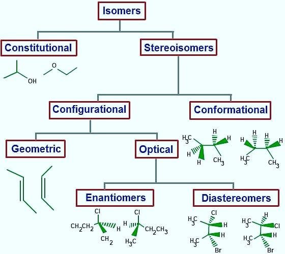
The branch of science that examines the bonding structures of atoms in the molecule and their arrangement in space in three dimensions is called stereochemistry. He studies how atoms are arranged in their relative positions in space. Isomerism is a discipline that studies the arrangement of molecules in space. Isomers are two or more different chemical compounds that have different bonding structures between their atoms but can be represented by the same molecular formula. These are compounds that have the same molecular formula but different arrangements of atoms. At the same time, the chemical properties of isomer compounds are also different.
– Basic classification of isomers
Stereoisomers
The covalent bonds and functional groups of a biomolecule are very important in the function of that molecule. The three-dimensional form of the molecule is formed by atoms. Carbon compounds exist in the form of stereoisomers. Their atomic numbers are the same, but their arrangement in space is different, and they also show different physical properties. Molecular interrelationships between biomolecules are invariably stereospecific, which creates specific stereochemistry in the molecules. The arrangement of atoms is called configuration, these can be cis, trans configurations, or configurations of optical stereoisomers. These are referred to as three-dimensional structures in stereochemistry. A carbon atom is asymmetric with 4 different atoms or groups. A molecule containing one or more asymmetric carbons is also asymmetric.
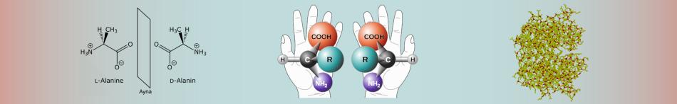
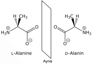
Enantiomers cannot be converted to each other. This is because enormous energies are required to break the covalent bonds and displace the atoms. Enantiomers have the same chemical properties as long as they are not in non-chiral environments. For example, there are two enantiomers of alanine, an amino acid, and their physical properties such as melting point, boiling point and solubility are the same, but if the enantiomers are mixed, the physical properties of the product such as melting, freezing point and solubility will be different, but their chemical properties will not change. If the ratio of enantiomers in a mixture is desired, the chromatographic and spectroscopic properties change by showing an externally asymmetric effect. Thus, the enantiomers act differently from each other and their analysis is done.
– Molecular structures of L- and D- Alanine (Eren et al. 2017).
Enantiomers
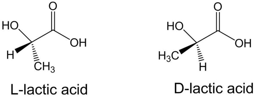
It is an asymmetric mirror image of the optically active substance (chiral substance) and has two enantiomers. It is formed by the bonding of the chiral molecule to four different groups or the carbon group of the atom with σ-bonds. Although the carbon atom is the asymmetric centre of the molecule, such molecules have a pair of stereoisomers with different spatial structures. They do not conflict with mirror images. Their optical rotation angles are the same in numbers but opposite in sign. The melting and boiling points, densities, and all physical properties of the two symmetric enantiomers are similar. Their chemical properties are the same in non-chiral environments, they react at a similar rate and form to form similar substances. Optical rotation angles are the same numerically but opposite in sign. It is possible to separate them from each other with advanced chemical analysis methods. Since most living molecules are enantiomers, there is an obvious difference due to the effects of the two symmetric enantiomers on living things, including humans.
– Enantiomeric forms of lactic acid (Casalini et al. 2019).
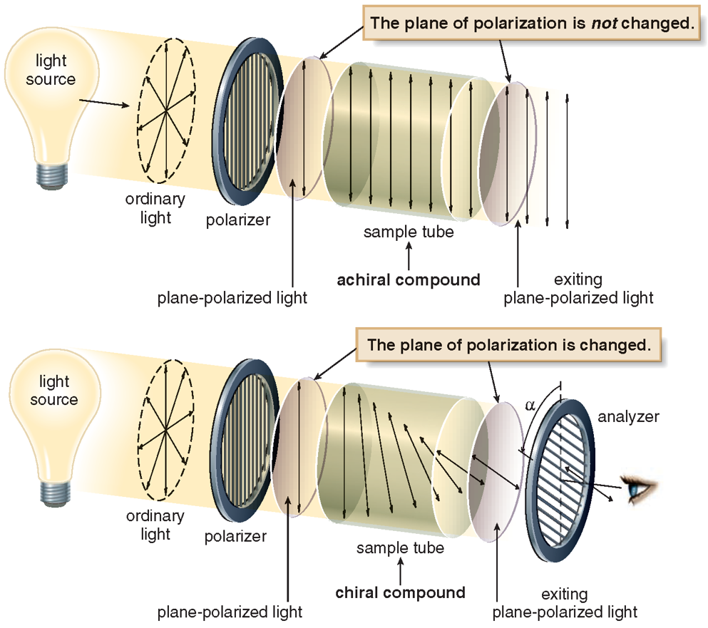
One of the physical properties of enantiomers is that the polarized beam has the ability to rotate the vibrational plane. For this reason, enantiomers are also called optical isomers. One of the optical isomers rotates the propagation of the polarized beam to the right, and the other to the left. Normal light emanating from a single source is a vibratory phenomenon that oscillates (spreads) in all directions and in all planes. Wave motion follows a path perpendicular to the direction of the light. Polarized light is light that vibrates in a single perpendicular plane and is free of wave vibrations. Enantiomer (symmetric) molecules make a definite spin when they encounter light, and when their mirror images are in the opposite direction, the spin is balanced and the spin of the light is reset. The net rotation of a single non-mirrored enantiomer in solution is not reset. Therefore, the rotation of polarized light is reset to zero in racemic mixtures. The specific rotation angles of optically active substances can be measured with a polarimeter. The enantiomer that rotates the vibrational plane of polarized light to the right is called dextrorotatory, or the enantiomer that rotates to the right, and the enantiomer that rotates to the left is called levorotatory or left-handed.
Chirality
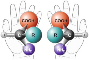
Chirality is a geometric property. If we explain this with an example from daily life if an item does not coincide with its mirror image, it is chiral, and if it coincides with its mirror image, it is not chiral. The characteristic of the chirality molecule is that the mirror image of the arrangement of the atoms of the molecule in space does not overlap. The chiral molecule has an asymmetric centre. Besides single chiral, there are chiral with more than one asymmetric centre. These are called multichiral structures. A molecule, however, may not exhibit chiral properties. Even if four different groups are attached to the sp3 hybridized carbon atom in a compound, it is chiral. If the asymmetric carbon atom is the centre of 4 different atoms or groups of atoms, it is called a stereogenic centre. Chiral compounds are asymmetric molecules due to the absence of an intramolecular symmetry plane and have two configuration isomers whose mirror images do not overlap each other. These two isomers are called enantiomers and these structures are enantiomeric to each other.
– Two enantiomers of a chiral generic amino acid
Nomenclature and properties of chirality
The R and S rules, or Chan-Ingold-Prelog (CIP), is used to name two enantiomers with opposite configurations. This naming is done within the framework of some rules. Simply put the smallest group is placed behind the stereogenic carbon atom and the remaining three groups are decided based on their order of precedence by looking at their right (R) or left (S) orientation. However, in the event that a decision cannot be made with this rule, and if the first bonded atom is the same, the atoms that continue in the second or third order are examined and a decision is made.
After passing through the chiral molecules, plane-polarized light ceases to be a plane, rotates and forms an angle. This is called optical activity. On one side the angle becomes +α° (clockwise) and on the other side, the angle becomes -α° (counterclockwise). For example, at the same concentration and experimental conditions (S)-carvone rotates plane-polarized light +10° clockwise, while (R)-carvone rotates plane-polarized light -10° counterclockwise.
– Interactions of chiral and achiral compounds with plane-polarized light
The biological significance of chirality
The existence of chiral molecules and the existence of molecules with stereogenic centres have also made great contributions to organic chemistry and other branches of organic chemistry.
Chirality is not required for biological activities to occur, but the enantiomeric activity of biologically active molecules with stereogenic centres is different. For example, it is found in biologically active substances such as food additives, fragrances and flavours, pesticides, herbicides and medicines. Living organisms contain largely chiral molecules in their components, and these chiral molecules play a dominant role in the mutual action of bioactive substances. All physical, chemical, and electrical differences experienced in living systems are perceived by free nerve endings or receptors with sensory receptors. Whatever their physiological function, the common feature of receptors is that they are chiral. Thanks to this feature, excitatory molecules behave enantioselectively during binding.
Many pharmaceuticals and chemical compounds used in disease treatment are composed of chiral enantiomers with the same physical and chemical properties in racemic structure. In the pharmaceutical industry, one of the enantiomers is desired curative, therapeutic, etc. While the other enantiomer may be inactive, it may have a beneficial effect or may cause harmful side effects. Since there is a need for chiral compounds of enantiomeric purity in the pharmaceutical industry, it is important to develop appropriate separation methods and establish new strategies in the purification processes of chiral molecules from racemic mixtures. Many optical resolution methods are used to separate racemic compounds. However, classical methods require a long process. This situation creates high costs, and a low amount of optically active compound is obtained in return, therefore it is not preferred in the pharmaceutical industry. Membrane-based chiral separation processes are economical compared to other separation processes due to their high efficiency and energy saving. In addition, this method is preferred because the scale-up step is easy.
Chiral molecules
Enzymes: Enzymes, which are chiral compounds, play a role in the realization of many reactions with their cofactors and coenzymes. They act as biocatalysts. Enzymes, with their enantioselectivity, are catalysts for asymmetric synthesis in oxidation-reduction reactions.
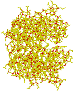
Amino acids: The functions of proteins depend on their interactions with other molecules through chiral structures. Homochirality of amino acids is necessary for the gene to specifically code for protein because L- and D-amino acids form three-dimensional protein structures. For example, the amino acids alanine and serine each have a stereogenic centre marked with an asterisk (*). In nature, they occur as natural enantiomers. Proteins, on the other hand, are actually a single enantiomer, despite having many chiral centres.
– A protein molecule and its stereogenic centres (red structures).
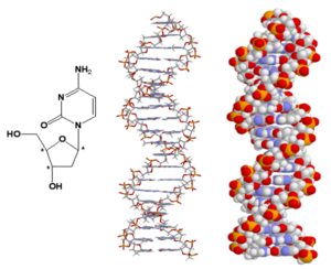
DNA: One of the basic building blocks of living things, the double-stranded DNA, in which genetic information is found, is also a chiral molecule. For example, the nucleic acid cytosine in DNA has three stereogenic centres indicated by an asterisk, and one can speak of chirality. Like proteins, DNA is a single enantiomer despite having many stereogenic centres.
– Cytosine molecule (far left) and DNA strands marked with three stereogenic central star symbols, which are chiral molecules.
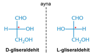
Carbohydrates: They are examined in three subgroups monosaccharides, disaccharides, and oligosaccharides. Carbohydrates are also in the class of chiral molecules.
– Stereogenic centres of D- and L-glyceraldehyde, one of the simplest monosaccharides, are indicated by *.
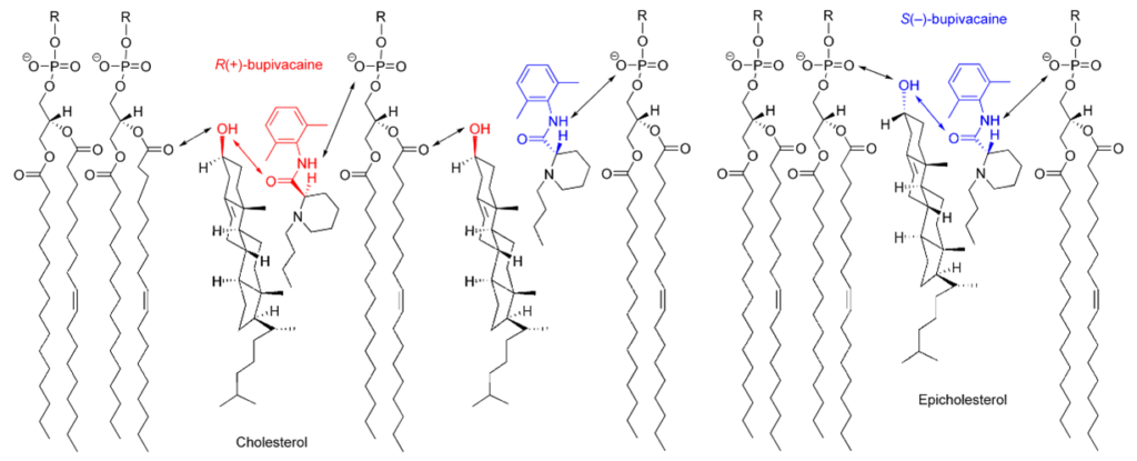
Lipids: Lipids are chiral molecules, which are the most important units of the cell membrane. He earned this title thanks to his optical activity. The enantiospecific interactions involved here change the properties of the cell, which can significantly affect the role of membrane-bound proteins.
– Some chiral lipid molecules in the cell membrane.
Pharmaceuticals: The importance of chirality in the pharmaceutical industry is an undeniable fact. The enantiomer, which is a mirror image of each other, behaves as two different compounds in the chiral environment, in addition to the difference between the directions of rotation of the vibrational plane of the polarized light. Therefore, their chemical properties in the chiral environment are also different. Since enantiomers have chiral properties in living organisms and structures, they can act in two different ways on these living organisms and structures. In other words, enantiomers can have opposite effects on each other. For example, (S)-(-)-propranolol has been described as a β-blocker in the treatment of heart disease, but its enantiomer (R)-(+)-propranolol acts as a contraceptive. Therefore, the enantiomeric purity of this compound is very important in clinical use. The concept of chirality is very common in pharmaceuticals. Pravastatin sodium for cardiovascular diseases, sertraline hydrochloride for central nervous system diseases, salmeterol for respiratory system diseases, clopidogrel bisulfate for haematology diseases, esomeprazole magnesium for digestive system diseases, potassium clavulanate as an antibiotic can be given as examples.
While 56% of the drugs used by the patients are chiral molecules, 88% are found as racemic mixtures. However, when using racemic drugs, it is necessary to use two layers of the racemic mixture in order to fully absorb the unit amount of active substance. For example, (R,R)-chloramphenicol shows antibacterial properties, while (S,S)- chloramphenicol shows inactive properties. Therefore, using racemic mixtures is uneconomical as it wastes half of the resources. Therefore, a single enantiomer always shows more biological activity than a racemic mixture.
– Two enantiomers of chloramphenicol.
As a result, chiral molecules are of great importance for living things. After the discovery of the concept of chirality, studies on naming them were made easier in the expression of these molecules and the understanding of the structure of chiral molecules was facilitated. The right or left orientation of chiral molecules is the factor that changes the way it acts. This is due to the fact that the two enantiomers act differently from each other. Contributing to the formation of the basic building blocks of living things is one of the most important effects of chirality. The contribution of chirality to the pharmaceutical industry is great. Almost half of the drugs on the market are chiral and approximately 50% of them are enantiomer mixtures. From the past to the present, many chiral drugs have been produced, and while most of them have been beneficial, some have been harmed. Chirality is a concept that provides convenience in the field of drug production, if a solution is found for these damages and if the harmful enantiomers can be prevented from showing side effects in the living thing. Biological compatibility, metabolism rate, metabolites, secretion, potency and receptor selectivity, transporters and enzymes, and toxicity of the two enantiomers of chiral drugs can be different. The use of single enantiomer drugs can potentially lead to easier pharmacokinetics and more selective pharmacological profiles, despite reduced drug interactions and different metabolism rates of different enantiomers. Single isomer chemicals can be preferred to racemic chemicals because they are more effective, use less quantity and are more economical in terms of the production process.
This article is a part of the homework written by İremgül Çelikyurt and Melis Tarkan, our Organic Chemistry students for the Fall Semester of the 2020-2021 Academic Year.
References
Alija K. 2016. Biyolojik Aktif Bileşiklerde Kiralitenin Önemi. Lisans Araştırma Projesi. T.C. Ankara Üniversitesi Eczacılık Fakültesi, Farmasötik Kimya Anabilim Dalı.
Cheung D. 2007. Teaching Chemistry Through The Jigsaw Strategy. Quality Education Fund, Hong Kong. 2-5.
Çakmak R. 2008. PirKLe –Tip Kiral Kolon Kromotografisi Yöntemiyle Biyolojik Öneme Sahip Kiral Aminlerden (±)-Β-Metilfeniletilamin’in Rezolüsyonu. Yüksek Lisans Tezi. T.C. Harran Üniversitesi Fen Bilimleri Enstitüsü.
Çetin A. 2010. Kiralite ve Biyolojik Aktivite. 24. Ulusal Kimya Kongresi, Zonguldak Karaelmas Üniversitesi. Erişim adresi: http://kimyakongreleri.org/2010/2010-701.pdf; Erişim tarihi: 12.11.2020
Demelezi V. 2018. Tetraoksokaliks[2]Aren[2] Triazin-Bazlı Kiral Bileşiklerinin Sentezi Ve Enantiyoselektif Reaksiyonlarda Katalizör Olarak Kullanımı. Yüksek Lisans Tezi. T.C. Necmettin Erbakan Üniversitesi Fen Bilimleri Enstitüsü.
Gal, J. 2006. Chiral Drugs from a Historical Point of View. In Chirality in Drug Research (eds R. Mannhold, H. Kubinyi, G. Folkers, E. Francotte and W. Lindner). https://doi.org/10.1002/9783527609437.ch1
İnaki M, Liu J, Matsuno K. 2016. Cell chirality: its origin and roles in left–right asymmetric development.Phil.Trans. R. Soc. B371: 20150403. http://dx.doi.org/10.1098/rstb.2015.0403
Karaküçük A. 2006. Kimyasal ve Biyoteknolojik Yöntemlerle Kiral Yapıların Sentezleri. Doktora Tezi. Selçuk Üniversitesi Fen Bilimleri Enstitüsü.
Lalitha S., Sampath Kumar A., Stine K. J., Covey, D. F. 2001. Chirality in Membranes: First Evidence that EnantioselectiveInteractions Between Cholesterol and Cell Membrane Lipids Can Be a Determinant of Membrane Physical Properties. Journal of Supramolecular Chemistry, 1(2): 53-61
Serpek, B. 2015. Organik Kimya. Nobel Akademik Yayıncılık
Smith JG. 2010. Organic Chemistry, 3rd Edition, McGraw-Hill.
Smith JG. 2012. General, Organic, & Biological Chemistry 2nd Edition, McGraw-Hill.
Our review article entitled “Biochemical Significance of Biomaterials Based on the Chitin-Chitosan Axis”, co-authored with my 2nd-year students Tamar Faraj and Mariam Ajam from NEU Faculty of Veterinary Medicine, was published in the journal “Acta Scientific Gastrointestinal Disorders (ISSN: 2582-1091)”.
I would also like to thank Ahmet Özer Şehirli PhD Assoc. Prof., Sevgi Gençosman DVM and Deniz Ceylanlı DVM, who contributed to the planning, literature review, writing, review and finalization of the article and with whom I enjoyed working and producing.
In our study, we conveyed the biological properties of the biomaterial called Chitosan, which is obtained from the polysaccharide called Chitin, which is the most common in nature and an important component of the exoskeletons of arthropods, and current approaches regarding its use in the field of health and my views in this direction from a biochemical point of view.
Nuclear Magnetic Resonance spectroscopy (NMR) is used to define organic compounds. Carbon (H) and Hydrogen (H) are high in organic molecules. NMR is used for structural analysis because it provides convenience in understanding the molecular structure; this knowledge contributes to scientific development.
NMR spectrum interpretation is at the forefront, and it is essential to understand the logic of spectrum measurements and act according to this logic while interpreting. In an atom and its properties, the electrons around the nucleus rotate both around the nucleus and around itself. Still, the movements of protons and neutrons in the nucleus are not noted. That’s why we do not know much about the movement of the nucleus. The nucleus’ protons rotate around their own axis, just as electrons around their own axis. As a result of this movement, the concepts of +1/2 and -1/2 are mentioned. Electromagnetic behaviour due to the spins of protons in the nucleus connects the proton, neutron and their electromagnetic interactions with each other with the logic of being destroyed or not. Like two electrons in an orbital, protons spinning in the opposite direction cancel each other’s electromagnetic effect. So the nucleus does not have a specific magnetic field.
NMR spectroscopy is primarily used by chemists, biochemists, and physicists who are working with complex nanoparticles. Biochemists use it for identifying complex molecules like proteins or intracellular metabolites. It has made a significant contribution to the medical area. Exploratory and diagnostic areas of medicine can exploit the NMR. The medical area is also used for magnetic resonance imaging (MRI).
History of NMR Spectroscopy
The first NMR signal was observed by two separate groups of physicists in 1945. Felix Bloch and Edward Mills Purcell were awarded the Nobel Prize in Physics in 1952 for their discovery. In the same year, NMR spectroscopy was used in molecular structure determination in chemistry. In 1953, the first NMR devices were produced. After 1970, devices with high discrimination power and sensitivity started to be made. Thanks to his work on developing high-resolution NMR spectroscopy, the scientist named Richard Robert Ernst won the Nobel Prize in Chemistry in 1991.
Felix Bloch
Edward Mills Purcell
Richard Robert Ernst
A chemist/biophysicist named Kurt Wüthrich won the Nobel Prize in Chemistry in 2002, thanks to his method for the investigation of biological macromolecules by NMR spectroscopy. Paul Christian Lauterbur and Sir Peter Mansfield were awarded the Nobel Prize in Physiology or Medicine in 2003 for their work in the NMR imaging field.
NMR spectroscopy has been used in chemistry, physics, biochemistry, pharmacy, and medicine to examine the structures of molecules.
Kurt Wüthrich
Sir Peter Mansfield
Paul Christian Lauterbur
NMR Spectroscopy
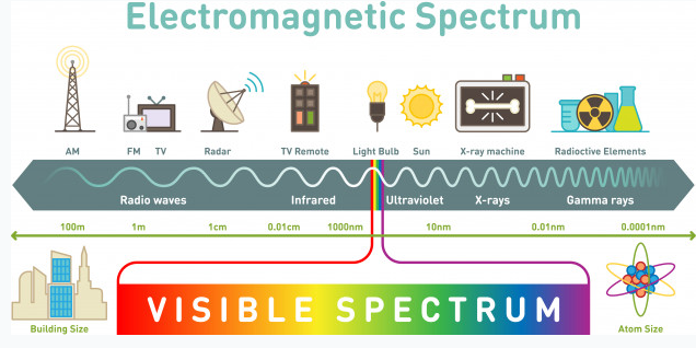
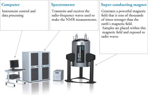
NMR spectroscopy is an illumination method based on the absorption of electromagnetic rays in the radio frequency field by atomic nuclei in a molecule placed in a strong magnetic field. NMR spectroscopy is a technique used in understanding the structure of molecules in the field of chemistry. Hydrogen-containing groups in the molecule and neighbouring groups can also be detected using this method. If evaluated together with the results obtained by other spectroscopic techniques, the structure to be illuminated can be easily reached. In UV and IR spectroscopy, the molecule’s functional groups and the percentages of C, H, O, N, and S atoms in the elemental analysis are determined. NMR spectroscopy gives information about the skeleton of the molecule. Other spectroscopic methods deal with electrons, but NMR spectroscopy deals with the nucleus. NMR requires a strong magnetic field and radio waves, which are long-wavelength rays of the electromagnetic spectrum. NMR spectroscopy does not disrupt the molecule, and analysis samples can be used repeatedly as in UV and IR spectroscopy.
Every atom whose atomic number or mass number is odd has a nuclear spin. The nucleus rotates around itself and is electrically charged creating its magnetic field. Rotating protons behave like bar magnets when placed in an external magnetic field. Rotating protons’ own magnetic fields go either in the same direction with the outer field or in the opposite direction. With the absorption of a photon with a certain amount of energy, the direction of the proton field can change. The energy difference between the two states is directly proportional to the strength of the magnetic field. Protons are surrounded by electrons that protect them from the external magnetic field. Rotating electrons create an exciting magnetic field opposite the external magnetic field and reduce the external field’s influence.
Charged particles rotating around their own axis create electrical and magnetic fields. When these atoms are placed in a stronger magnetic field, two magnetic spins called +1/2 and -1/2 occur. Depending on the direction of the outer magnetic field, the magnetic field due to the atom’s own spin is either added or removed. Thus, the higher and lower energy nucleus can be found.
The small magnetic field in the core and the large applied magnetic field cause the difference in the results of subtraction and addition to be small. This difference changes with the applied external magnetic fields and becomes zero if the magnetic field is not applied. NMR spectroscopy is based on trying to equalise the difference from the external magnetic field with radio frequencies.
Magnetic Properties of the Nucleus and the Basis of NMR Spectroscopy
Some atomic nuclei act like magnets rotating around themselves forming the basis of NMR spectroscopy. Atomic nuclei are positively (+) charged. The nucleus rotates around itself, and the (+) load moves in orbit around the axis, this is the spin motion. Because the nucleus rotates around itself, it also has angular momentum. A dipole and a magnetic field arise from the spin motion. The size of the dipole is called the nuclear magnetic moment (μ), and the angular momentum of the charge is called the spin quantum number (I). To study the NMR of an element, the magnetic moment must be non-zero (μ≠0), and the spin quantum number must be greater than zero (I>0). The spin quantum number varies according to the number of protons and neutrons in the nucleus. The spin number can be 0, 1/2, 1, 3/2, 5/2. If I=0, there is no spin. Protons and neutrons have their own spins, and their sum gives the number of spins in the nucleus. Isotopes of an element have different spin quantum numbers.
Proton NMR Spectroscopy
NMR spectroscopy, like other spectrophotometers, examines samples in dilute media. First, the sample solution to be NMR is taken into a glass tube of 5 mm in diameter and 15 cm in size and placed in a strong magnetic field. Then the radio frequency is sent to the sample. After the sample emits the radio frequency it has absorbed, the detector measures the re-emitted frequency. This change is related to the externally applied magnetic field, and the applied magnetic field must be kept constant throughout the measurement.
Components of NMR instrument (Alqaheem Y, Alomair AA: Microscopy and Spectroscopy Techniques for Characterization of Polymeric Membranes. Membranes (Basel) 2020; 10.)
NMR instrument composition
Protons behave differently from each other, which is explained by the electron density around them. Although the proton’s magnetic properties are equal, it is accepted that they will differ in the magnetic environment due to the electron density around it, thus making it easier to understand the spectrum that NMR spectroscopy will give. This difference is minimal compared to the magnetic field and is expressed using ppm (parts per million). Generally, the frequency scale is used instead of magnetic field differences in NMR. The proton resonances of organic molecules are between 0-12 ppm. Highly sensitive devices and systems are required to examine this small range.
The Determination of theStructure of Membrane Proteins using NMR Spectroscopy
Membrane proteins are a part of the biological membranes. They are branched into several types. They can penetrate the cell or be on its surface and have a temporal interaction. Membrane proteins have an important role in the medical area. They are the target structures of the drug. Besides, they have a crucial impact on human and animal diseases. Membrane proteins have the capability of mutating and truncating. While these procedures, several diseases can occur in both humans and animals.
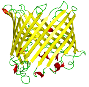
Membrane proteins have a wide range of characteristics, such as being a receptor and interacting with hormones. They can reveal as enzymes and bind to the receptors. Some of them are membrane transporter proteins and impact the transportation of small molecules, macromolecules, and some ions. Their secondary structure is composed mainly of β-barrel, a β-sheet consisting of a first and last strand bonded with hydrogen bonds.
18-strand β barrel.
β-barrels mostly can dissolve in water. Strands of the beta barrels contain polar and nonpolar amino acids which lead the protein to have hydrophobic and hydrophilic sides; mostly the hydrophilic part interacts with the solvent because it is a settled surface. The hydrophobic part plays inside of the protein molecule. In proteins containing beta barrels, the hydrophobic side is towards the exterior. They interact with the lipids under the vicinity of it, which surround the protein’s exterior.
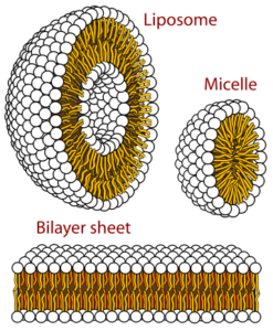
The structures formed by phospholipids in aqueous solutions. Micelles are single-chain lipids.
GPCRs (G-protein coupled receptors), membrane proteins, are cell signal transportation receptors. The 3D structure of these proteins is determined by NMR spectroscopy. In a region made out of lipids, we can observe how membrane proteins’ motions are reflected. It can be observed in a small area or a large area. As the scientists are supposed to dissolve the substance to mark it on the NMR spectroscopy, they use detergents to dissolve, to denature proteins.
While solubilizing the proteins using detergents, some substances occur—for example, micelles and isotropic bicelles. Micelles are the strewn surface-active molecules’ flocculation. Isotropic bicelles are for the reconstruction of membrane proteins. They patch the proteins with lipid bilayers. Detergents are unnatural substances, so they cause the risk of subverting the protein structure. You can most clearly study the proteins when they are in a phospholipid environment.
The helical membrane proteins can be observed at a high resolution under the bilayers’ favour. They are in an active and immobilised state. On the NMR, substances are well determined in a high-resolution solid state. Bacteriorhodopsin is the first determined helical membrane protein under NMR spectroscopy. It was solubilised with octyl glucoside, which is a type of detergent. Other than NMR spectroscopy, X-rays have also contributed to the determination of membrane proteins. This type of protein was first observed under x-ray diffraction. It was observed during a photosynthetic reaction. The product of this reaction was Rhodopseudomonas viridis (a kind of bacteria), and it was solubilized in a powerful detergent, N, N-dimethyl dodecyl amine N-oxide. The detergent is composed of amphiphilic (both have the characteristic of being hydrophobic and hydrophilic) compounds.
Carbohydrate-Protein Interactions
NMR can identify carbohydrate-protein interactions with the resolution in solvents. This interaction is necessary for the virus-to-cell and cell-to-cell connection. Continuous interaction is provided by the carbohydrate-protein mutual effect for an infection or adhesion event. Even in viruses and pathogens, the outside of the cell is surrounded by glycans. Recognisance and biological transactions are instructed by the mutual effect of the glycans and protein receptors. A variety of molecules can have a mutual effect on carbohydrates. It happens in the vicinity of a mutual effect between L-selectins and glycans terminated by sialic acid.
Influenza infection requires binding to carbohydrates. Hemagglutinin cells (influenza virus) have to be connected to the Siaα-6Gal. The adhesion and growth of tumour cells are supported by β-lactosamine’s mutual effect, which includes galectins and glycans. There are several approaches to NMR. It can be protein-based or ligand-based. A ligand is a complex compound that contains attached biomolecules. If it is protein-based, it will have different solubility characteristics from other solutions. High-resolution attainability requires labelling the protein with C and N stable isotopes. By this labelling procedure, the size will be in the range of 10-25 kDa, which is in NMR standards. There are some protein-detecting methods. In the protein’s 3D structure, the scientist must map the linkage regions of the carbohydrate onto it. The linkage regions are determined by the correlation spectra of H and N atoms. Ligands of carbohydrates must be defined because they are specified from various proteins. In terms of the NMR method (methods of protein detection), it is crucial to compare the proteins in their free state and bound to the carbohydrate state. The labelled protein quantity is dependent on several topics. The first one is the instrumentation of NMR, and the second one is the spectrometer’s available time.
NMR Spectroscopy for the Assignation of Unsaturated Fatty Acids
Fatty acids (FAs) are usually identified by gas chromatography (GC). FAs have to be transformed into methyl esters to assign them to GC. There are saturated and unsaturated FAs. Unsaturated FAs (UFAs) carry one, two or more double bonds and are critical components of animal fats and vegetable oils. UFAs having two or more double bonds are referred to as Polyunsatured FAs (PUFAs). There is a method based on the carbon NMR To determine these bonds’ percentages. Besides, scientists also use hydrogen to quantify UFAs.
Chemical Shift
NMR signals change depending on the magnetic field and radio frequencies. Therefore, radiofrequency change is studied in a standard magnetic field or in a magnetic field change under a standard radio frequency. This problem is avoided by adding a standard substance during NMR measurements. This substance should not affect the hydrogen atom’s electron density with its electronegativity much, and it is tetramethylsilane (TMS) without a solubility problem. All the hydrogens of TMS are equal and at the same point. This point is referenced (point O). The interpretation of where other hydrogen atoms come out according to this point is called the chemical shift. Its unit is ppm, and the symbol represents it. During NMR measurements, it is preferred that the molecules whose spectrum is to be taken be diluted. The presence of hydrogen atoms in the solvent causes the problem of intensity. Some solvents do not contain hydrogen, and generally, not all substances have good solubility. To solve this problem, deutero structures that are not affected by the magnetic field are used. The most common and first tried solvent is deutero chloroform. Solvents such as deutero water, ethyl alcohol, and dimethylsulfoxide are also widely used.
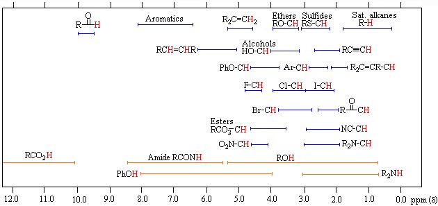
Proton chemical shift values
Before making the NMR spectrum interpretation, the electron density around the hydrogen atom must be known from a rough perspective. Compared with TMS, the electron density around a hydrogen atom changes depending on the electronegativity and shielding effect of the atoms to which it is attached. If we accept TMS=0 ppm, Si electronegativity is less than C electronegativity, so the electron density in hydrogen atoms bound to carbon atoms will be less than TMS and shielding less. Thus, we see that radio frequency signals reach the protons in the nucleus and return; thus, excitation and resonance will be easier. It requires less magnetic field, so the ppm gradually increases from 0. Therefore, C-H’s are attached to the carbon atom in molecules that do not contain electronegative, such as 0-1. The chemical shift of hydrogen atoms with low electron density and shielding around them, such as the RCOOH acid proton, is as large as 11–12 ppm. Although there are tables for proton chemical shift values, it is often possible to approximate peaks and locations. It is not always necessary to look at this crowded picture, but it may be necessary with some complex and large molecules.
In the NMR spectrum, the signal intensity depends on the substance concentration. Dilute solutions give weak signals. If the concentration increases, the peak intensity increases. If we take the peaks of different substances at the same concentration, we see that the peak intensity depends on the equivalent hydrogen number. If we take the equivalent concentration of benzene containing 6 equivalents of hydrogen and cyclohexane containing 12 equivalents of hydrogen and measure the NMR spectrum, the NMR spectrum peak intensity of the cyclohexane is twice that of benzene. When these results are combined with the knowledge of how many hydrogen atoms it contains and chemical shift, it makes it easier to understand where the hydrogens of the molecule come out. However, there are still some problems with knowing how many hydrogens each peak equals. However, hydrogen numbers can be calculated from the ratio of peaks to peaks, but instead of exact numbers, they are found in coefficients relative to each other. It is possible to estimate approximate values with this integration method, similar to a simple molecular formula calculation.
In conclusion, NMR spectroscopy has contributed to the determination of various complex molecules like proteins, carbohydrates, enzymes, intracellular metabolites, receptors, and transporters. Understanding these structures by scientists leads to the development of science. Complex molecules are essential to understand various diseases, both in humans and animals. Although NMR is complicated for us, it is a beneficial thing today. That’s why we should try to grasp a complex subject’s logic rather than make it difficult in our minds. The blessings of NMR are countless. It is used in many industries, such as polymer research, synthetic chemistry, petrochemistry, biochemistry, textiles, food, paint, medicine, and agriculture. We can achieve many things using NMR, such as the compound’s nature, structure shape and bonding structure, mixture components, atomic composition, molecular weight and formula, polymer composition and arrangement, and molecular motion. No matter how boring its theory may sound, we do not know how correct it is to call something ‘boring’ that we use somehow.
This article is a part of the home assignment written by Ada Begüm Ögel and Emir Kerem Demiroğlu, two of our 2020-2021 Academic Year Fall Semester Organic Chemistry students.
References
Alqaheem Y, Alomair AA: Microscopy and Spectroscopy Techniques for Characterization of Polymeric Membranes. Membranes (Basel) 2020; 10.
Aletli Analiz Yöntemleri: Nükleer Manyetik Rezonans (NMR) Spektroskopisi [http://web.hitit.edu.tr/dersnotlari/gokcemerey_13.10.2015_7A4L.pdf]
Shulman GI, Alger JR, Prichard JW, Shulman RG: Nuclear magnetic resonance spectroscopy in diagnostic and investigative medicine. J Clin Invest 1984; 74:1127–1131.
Bewley CA, Shahzad-ul-Hussan S: Characterizing carbohydrate-protein interactions by nuclear magnetic resonance spectroscopy. Biopolymers 2013; 99:796–806.
Wider G: Structure determination of biological macromolecules in solution using nuclear magnetic resonance spectroscopy. Biotechniques 2000; 29:1278–82, 1284–90, 1292 passim.
Campagne S, Gervais V, Milon A: Nuclear magnetic resonance analysis of protein-DNA interactions. J R Soc Interface 2011; 8:1065–1078.
Simpson MJ, Hatcher PG: Determination of black carbon in natural organic matter by chemical oxidation and solid-state 13C nuclear magnetic resonance spectroscopy. Org Geochem 2004; 35:923–935.
Opella SJ, Marassi FM: Structure determination of membrane proteins by NMR spectroscopy. Chem Rev 2004; 104:3587–3606.
Miyake Y, Yokomizo K, Matsuzaki N: Determination of unsaturated fatty acid composition by high-resolution nuclear magnetic resonance spectroscopy. J Am Oil Chem Soc 1998; 75:1091–1094.
Chalbot M-CG, Kavouras IG: Nuclear magnetic resonance spectroscopy for determining the functional content of organic aerosols: a review. Environ Pollut 2014; 191:232–249.
Zia K, Siddiqui T, Ali S, Farooq I, Zafar MS, Khurshid Z: Nuclear Magnetic Resonance Spectroscopy for Medical and Dental Applications: A Comprehensive Review. Eur J Dent 2019; 13:124–128.
The incidence of non-Hodgkins Lymphoma (NHL) or Malignant Lymphoma (ML) in dogs is reported to be more than 24 per 100,000. Advances in the diagnosis and treatment of ML in dogs not only improve the quality of life of animals but also enable better models in veterinary comparative oncology.
Thymidine kinase (TK) is an intracellular enzyme that plays an important role during pyrimidine synthesis. TK activity increases markedly in the G1-S phase, especially during cell division, and decreases rapidly in the G2 phase. Therefore, high extracellular TK activity reflects high DNA synthesis and cells that die during cell division. Hematopoietic system malignancies are characterized by high cell proliferation. Studies in the veterinary field have shown that serum Thymidine kinase activity is an important marker in the diagnosis, prognosis and monitoring of treatment efficacy in leukaemia, multiple myeloma and malignant lymphoma.
TK activity has been used for years in the diagnosis, prognosis and treatment follow-up of hematopoietic tumours in human oncology, and the first study in the veterinary field was conducted by Nakamura et al. conducted in 1997 in Lymphoma, leukaemia, non-hematopoietic tumours (breast tumour, mastocytoma, anal sac tumour, malignant histiocytosis) and healthy dogs. After the analysis, TC activity in dogs with Lymphoma and Leukemia was significantly increased compared to healthy dogs; in dogs with non-hematopoietic tumours, it was found to be at the same level as healthy dogs. Again in the same study, it was determined that TC Activity is important in the follow-up of the treatment in the analyzes performed before the treatment, at the stage of the disappearance of clinical symptoms, and at the relapse stage.
In another study conducted by Prof. Dr. Hendrik von EULER et al. in dogs diagnosed with ML between 1999-2003, it was reported that TK Activity can be used as a strong marker in the diagnosis of ML disease, especially in determining the prognosis and predicting clinical disease before a recurrence in dogs undergoing chemotherapy. Serum TK Activity was found to be 2 to 180 times higher in dogs with ML disease than in healthy dogs. It was determined that TC activity decreased to normal values in dogs that responded to treatment and whose cancer symptoms disappeared (complete remission), and TC activity increased again before recurrence. In the same study, it was determined that TC activity was correlated with the clinical stages of the disease.
Similar studies have been carried out in cats in recent years, along with studies in dogs. The first study on cats was conducted on a total of 171 cats in the UK and Sweden, published in 2012 and also included in our partner laboratory, Dechra Specialist Laboratories. Of the cats included in the study, 49 were healthy, 33 had lymphoma, 55 had the inflammatory disease, and 34 had non-hematopoietic neoplasia. At the end of the study, it was determined that the serum TC activity was significantly higher in cats with lymphoma compared to the others, and it was reported that high TC activity would strengthen the diagnosis of lymphoma.
Thymidine kinase activity with recent studies;
In the diagnosis of lymphoma and leukaemia together with other clinical and laboratory findings,
In evaluating the prognosis,
In the evaluation of chemotherapeutic success with analyzes performed before, during and after treatment,
Monitoring chemotherapy and identifying relapse cases before they form,
It has been used successfully in distinguishing clinical worsening in patients receiving chemotherapy treatment.
References
Boyé, P. et al. (2019) ‘Evaluation of serum thymidine kinase 1 activity as a biomarker for treatment effectiveness and prediction of relapse in dogs with non-Hodgkin lymphoma.’, Journal of veterinary internal medicine, 33(4), pp. 1728–1739. doi: https://doi.org/10.1111/jvim.15513.
Bryan, J. N. (2016) ‘The Current State of Clinical Application of Serum Biomarkers for Canine Lymphoma.’, Frontiers in veterinary science, 3, p. 87. doi: https://doi.org/10.3389/fvets.2016.00087.
von Euler, H. et al. (2004) ‘Serum thymidine kinase activity in dogs with malignant lymphoma: a potent marker for prognosis and monitoring the disease.’, Journal of veterinary internal medicine. United States, 18(5), pp. 696–702. doi: https://doi.org/10.1371/journal.pone.0137871.
Kayar, A. et al. (2018) ‘Clinical features, haematologic parameters, blood serum biochemistry results and thymidine kinase activity of dogs affected by malignant lymphoma in Turkey’, Japanese Journal of Veterinary Research, 66(4), pp. 227–238. doi: https://doi.org/10.14943/jjvr.66.4.227.
Larsdotter, S., Nostell, K. and von Euler, H. (2015) ‘Serum thymidine kinase activity in clinically healthy and diseased horses: a potential marker for lymphoma.’, Veterinary journal (London, England : 1997). England, 205(2), pp. 313–316. doi: https://doi.org/10.1016/j.tvjl.2015.01.019.
Nakamura, N. et al. (1997) ‘Plasma thymidine kinase activity in dogs with lymphoma and leukemia.’, The Journal of veterinary medical science. Japan, 59(10), pp. 957–960. doi: https://doi.org/10.1292/jvms.59.957.
Selting, K. A. et al. (2016) ‘Thymidine Kinase Type 1 and C-Reactive Protein Concentrations in Dogs with Spontaneously Occurring Cancer.’, Journal of veterinary internal medicine, 30(4), pp. 1159–1166. doi: https://doi.org/10.1111/jvim.13954.
Taylor, S. S. et al. (2013) ‘Serum thymidine kinase activity in clinically healthy and diseased cats: a potential biomarker for lymphoma.’, Journal of feline medicine and surgery. England, 15(2), pp. 142–147. doi: https://doi.org/10.1177/1098612X12463928.
Blood creatinine concentration is measured for purposes such as diagnosis of kidney failure, determination of its stage, follow-up of treatment, and evaluation of prognosis.
In addition, from time to time, urine creatinine concentration is measured and evaluated alone or in combination with other test parameters (such as protein). The only way to make these measurements is to use specific analytical methods. One of the commonly used methods is the Jaffe reaction. The creatinine concentration is determined from blood and urine samples by the Jaffe reaction, which is a colorimetric method1.
132 years ago (1886) Max Jaffe (1841-1911) discovered that creatinine reacts with picric acid in an alkaline environment and explained this by publishing his article “Über den Niederschlag, welchen Pikrinsäure in normalem Harn erzeugt und über eine neue Reaction des Kreatinins”2. The article describes this reaction and the nature of the precipitate formed. Jaffe’s discovery was a turning point. As a result of this study, the method of measuring creatinine concentration, which has become extremely popular and defied time, was born.
Over time, Jaffe’s name became synonymous with clinical creatinine testing, although his article later became the permanent method and the principle of further studies. At the beginning of the twentieth century, Otto Folin (1867-1934), taking up the research of Max Jaffe, developed a colorimetric method for measuring the concentration of creatinine in blood and urine3 and made it into modern biochemistry analysis.
Although there are more specific analytical methods4 today, this unique test is still used as the preferred method due to its simplicity of implementation, speed, compatibility with automated analyzers, and cost-effectiveness. Besides, the Jaffe reaction is the oldest test method used in clinical laboratories.
References
Delanghe JR, Speeckaert MM. Creatinine determination according to Jaffe – What does it stand for? NDT Plus. 2011;4(2):83-86. doi: http://doi.org/10.1093/ndtplus/sfq211
Jaffe M. Ueber den Niederschlag, welchen Pikrinsäure in normalem Harn erzeugt und über eine neue Reaction des Kreatinins. ZPhysiolChem. 1886. doi: https://doi.org/10.1515/BCHM1.1886.10.5.391
Calcium (Ca) is one of the macro elements that have great importance in both animal and human metabolism.
In the regulation of total calcium (tCa) metabolism, mainly skin, liver, kidneys, bones and intestines at the tissue level; Parathyroid Hormone (PTH), Calcitonin (CT) and vitamin D take part at the molecular level. Calcium is the structural component of the skeletal system and has different and various functions in the organism. These include muscle contraction, blood coagulation, enzyme activity, neural stimulation, hormone release, secondary messenger, and membrane permeability.
The calcium ion concentration of the extracellular fluid in the body is vital and is always kept in balance. Parathyroid hormone (PTH), Calcitonin (CT) and Vitamin D contribute primarily to this balance. Apart from these, other hormones such as adrenal corticosteroids, estrogens, thyroxine, somatotropin and glucagon also contribute.
Calcium in plasma or serum is divided into 3 fractions. These:
Ionized or free calcium (iCa or Ca++) (≈56%)
Protein-bound calcium (mostly albumin) (≈34%)
Complex or chelated calcium (transports bound to various anions with small molecular weights-phosphate, bicarbonate, citrate, lactate) (≈10%)
iCa and complexed calcium form the dispersible fraction of calcium. This fraction may also be referred to as ultrafiltrate calcium as it passes through biological membranes. iCa is the most physiologically active fraction of serum calcium. iCa is responsible for functions such as bone homeostasis, nerve conduction, blood coagulation, Vitamin D and PTH secretion, activation of metabolic and digestive enzymes, and effective use of iron, and is also a sensitive marker of pathological conditions.
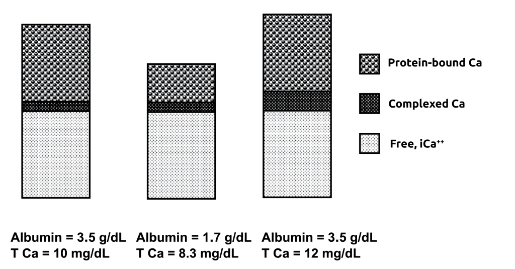
About 90% of protein-bound calcium is bound to albumin and the remaining 10% is bound to various globulins. Since approximately half of the calcium is bound to proteins, the evaluation of tCa depends on serum albumin and total protein values (Figure 1).
Figure 1.While iCa normally remains in a very narrow range, the tCa concentration is affected by either bound or complex calcium. In other words, the tCa concentration may differ depending on the change in protein-bound Ca or complex Ca fractions.
Traditionally, the assessment of an animal’s calcium status has been based on the assessment of its tCa concentration. The tCa concentration is assumed to be directly proportional to the biologically active fraction and iCa, the gold standard for the determination of calcium status. However, this assumption is not valid in a variety of clinical situations. It has been suggested that tCa can be corrected or adjusted according to albumin or total protein concentration to improve the diagnostic interpretation, especially in patients with hypoalbuminemia or hypoproteinemia, when iCa measurement is not possible. Also, changes in pH change the calcium fraction bound to albumin; therefore, the iCa concentration can also change without a change in tCa. This corrected or adjustable tCa is called corrected calcium (ctCa). Evaluation of ctCa is recommended, especially when the plasma albumin concentration changes.
Concentration measurement of tCa, albumin and total protein can be done easily with in-house and laboratory-type analytical devices. Generally, Arsenazo III, Bromcresol green and Biuret methods are used in this type of device, respectively.
The measurement of iCa concentration is made with devices with ion-selective electrodes (ISE). Such devices can be mobile or bench-type POC (point-of-care) devices (Figure 2), or they can be a component of automated biochemistry analyzers. Mobile-type devices are frequently preferred in clinics and are often costly, and it is recommended to compare the results with reference laboratory results; This process is recommended in doubtful cases or for quality control purposes from time to time.
When measuring iCa concentration, sample collection and processing should be done with the utmost care and attention. Samples should be collected in an anaerobic environment (to minimize carbon dioxide loss), transported in the cold chain and processed within a few hours (to minimize lactate production). tCa concentration measurements are relatively inexpensive, readily available, and more robust to sample transport variables. For these reasons, the measurement of total calcium is frequently performed and evaluated today.
As a result, it is reported that all three parameters can be used in monitoring the body’s Ca balance. The most important thing at this point is to understand what each parameter is, its variables and what could be misleading. The use of tCa as an indicator of Ca status, especially in hypoalbuminemia cases, tends to overestimate hypocalcemia and ignore normocalcemia; Using ctCa may result in overestimating normocalcemia and ignoring hypocalcemia. Therefore, it is recommended to evaluate Ca homeostasis with iCa concentrations instead of tCa or ctCa in hypoalbuminemia cases. Thus, it can be determined whether there is true hypocalcemia.
References
1-Caprita R, Caprita A, Cretescu I. Estimation of Ionized Calcium and Corrected Total Calcium Concentration Based on Serum Albumin Level. Anim Sci Biotechnol. 2013;46(1):180-184. 2-Danner J, Ridgway MD, Rubin SI, Le Boedec K. Development of a Multivariate Predictive Model to Estimate Ionized Calcium Concentration from Serum Biochemical Profile Results in Dogs. J Vet Intern Med. 2017;31(5):1392-1402. doi: https://doi.org/10.1111/jvim.14800 3-Bohn AA. Veterinary Hematology and Clinical Biochemistry. 2nd ed. (Thrall MA, Weiser G, Allison R, Campbell T, eds.). NJ, US: Wiley-Blackwell, John Wiley & Sons; 2012. 4-Payne RB, Carver ME, Morgan DB. Interpretation of serum total calcium: effects of adjustment of albumin concentration on frequency of abnormal values and on detection of change in the individual. J Clin Pathol. 1979;32(1):56-60. doi: https//doi.org/10.1136/jcp.32.1.56 5-Sharp CR, Kerl ME, Mann FA. A comparison of total calcium, corrected calcium, and ionized calcium concentrations as indicators of calcium homeostasis among hypoalbuminemic dogs requiring intensive care: Original study. J Vet Emerg Crit Care. 2009;19(6):571-578. doi: https//doi.org/10.1111/j.1476-4431.2009.00485.x 6-Toffaletti JG. September 2011 Clinical Laboratory News : Calcium. 2011;37(9):6-10.