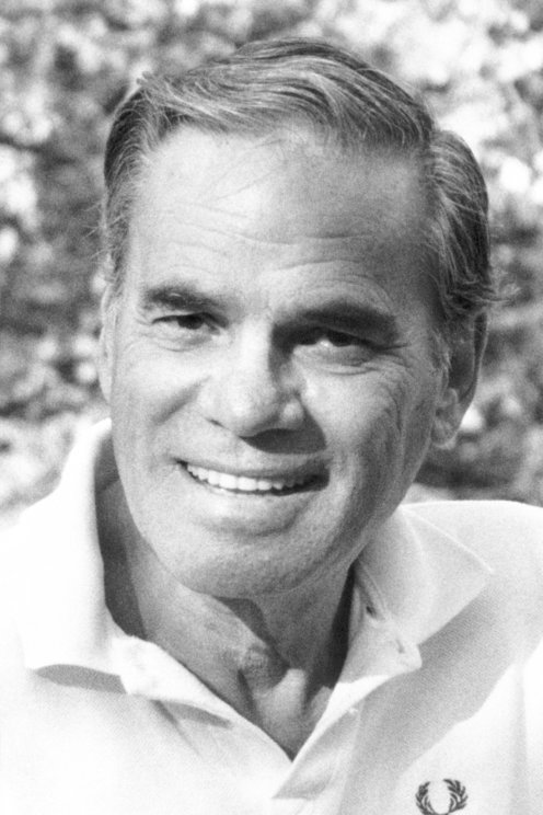
Martin Rodbell was a biochemist best recognized for discovering G-proteins. He received the Nobel Prize in Physiology or Medicine in 1994 for his studies on “G-proteins and their role in signal transduction in cells.”
Martin Rodbell, father of G-proteins, was born in Baltimore, Maryland, USA on December 1, 1925. After finishing his public school education there, he attended Johns Hopkins University in 1943. Even though he primarily studied Latin, Greek, German, and French, he became more interested in French because of his friends, and this interest affected the course of his educational life. While at university, he served in the navy in World War II and, as a Jew, believed that battling Hitler was a priority.
When he was serving as a navy radio operator on the Japanese front in the Pacific Ocean in 1944, he realised that the communication and experiences with different individuals under challenging circumstances had actually prepared him to become a scientist. When he returned from the war in 1946, he intended to continue his schooling at Johns Hopkins. While Rodbell was attracted to French literature, her father was eager to his attend medical school. The turning point in this process occurred when James Ebert recognised his passion for the philosophy of science and embryology and realised then he wanted to have a career in the biological sciences. To progress in the discipline of biochemistry, he stayed at Hopkins for another year and studied advanced chemistry courses, as suggested by Bentley Glass, one of the great professors in the biology department.
Once he met Barbara Ledermann in 1949, she brought with him a group of companions who were interested in a wide range of creative art. Rodbell, who married in 1950 and became a participant in the world of science and art, later relocated to Seattle and began working at the University of Washington. Working on lipid chemistry, specifically phospholipid metabolism, he learnt methods of examining the actions of phospholipases in ether solutions from his thesis advisor, Donald Hanahan. In 1954, he earned his PhD with his thesis on the biosynthesis of lecithin in rat liver. In the same year, the head of the University of Illinois’ Department of Chemistry, Dr Herbert E. Carter, contributed to the study of chloramphenicol biosynthesis. During his two years there, he also worked as a teacher, but he realized that he wanted to continue his research on the biochemistry of lecithin found in cell membranes.
Martin Rodbell accepted the position of biochemistry research assistant in Christian Anfinsen’s lab at the National Heart Institutes in Maryland in 1956.
Dr Edward Korn identified lipoprotein lipase, an enzyme that hydrolyzes chylomicron triglycerides, as a clearing factor. Rodbell, on the other hand, was interested in discriminating the nature of lipoproteins on the surface of chylomicrons and using the “fingerprint” method, he determined that there were at least five different proteins that will later be proven to have important roles in lipoprotein-containing diseases. When he decided to return to embryology research in 1960, he joined the University of Brussels. With the guidance of Jean Brachet, he observed the technique of X-ray films for recording the localization of tritium-labelled molecules in cells. After that, Dr In Peter Gaillard’s lab in Leiden, obtained specialized training in the use of cultured heart cells to differentiate the uptake of tritium-labelled chylomicrons.
Rodbell’s interest switched from the metabolic functions of lipid proteins to the effect of hormones (particularly insulin and glucagon) on individual cells while he was at the Arthritis and Metabolic Diseases Institute in the mid-1960s. Korn observed that the enzyme was found in adipose tissue, while Rodbell observed that collagenase digested and released fat cells. Following a meeting with Bernardo Houssay in 1963, he focused on the functioning mechanism of hormones on isolated cells and released his essay “Metabolism of Isolated Fat Cells” in 1964, which drew widespread attention in the field of endocrinology. After identifying that the mechanism of action of phospholipases mimicked the effect of the hormone on glucose utilization and protein synthesis, but that their action was limited to the surface membrane, he suggested that insulin-stimulating phospholipases could alter the structure of the surface membrane. All of these findings suggested that the insulin receptor is found on the surface of fat cells. Rodbell continued his studies on the hormone-sensitive fat cell, which he dubbed the “ghost,” by safeguarding the cell’s structural and metabolic features.
Rodbell greatly contributed to the understanding of the importance and function of G-proteins in 1969.
Following Earl W. Sutherland’s speech on hormones, activity drew the attention of many biochemists when he announced the hypothesis that the first messenger operates on the cell surface and triggers the “second messenger” mechanism, leading Rodbell to focus on the cyclic AMP paradigm. In 1967, when he travelled to Geneva, he collaborated with Torben Clausen on the effects of hormones on ions and amino acid translocations in “ghost” fat cells, concluding that hormones are pleiotropic agents. In 1968, while working with rat liver membrane cells, he believed the existence of a receptor for transducing and transmitting signals that trigger intracellular processes.
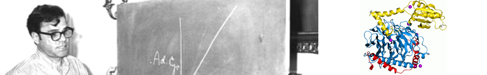
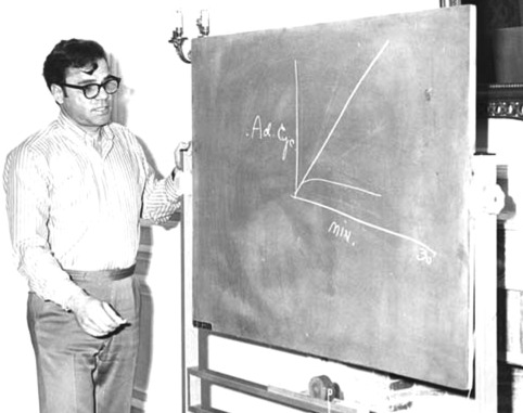
In 1969, he made a significant contribution to the understanding of the role and function of G-proteins by developing a system he dubbed “transmission of signal” for describing the components of cellular communication. Alfred G. Gilman and Rodbell during try to stimulate cells with adrenaline, discovered in 1970 that the main component of the cell transducer is a GTP (guanosine triphosphate)-dependent protein —Guanine nucleotide-binding protein/G-protein— that plays a role in impulse transmission. It is also known that when these molecules fail to perform their receptor functions adequately, they are linked to illnesses like diabetes, blindness, and allergies.
He was transferred to the National Institute of Environmental Health Sciences in 1985 after serving as Professor of Biochemistry at the University of Geneva from 1981 to 1983. Following his retirement, Rodbell and Alfred G. Gilman were awarded the Nobel Prize in Medicine and Physiology in 1994 for their independent work that led to the discovery of the G protein. After receiving the Nobel Prize, he went on to teach at high schools and universities. The Rodbell lecture series was initiated by the National Institute of Environmental Health Sciences, and Rodbell has spoken on the discovery process and its work at several of its conferences.
During his lifetime, Rodbell enables scientists from all over the world to share their opinions and experiences, and he defined his profession and life as “In many respects, my career and my experiences with people and events have been seamless in that I cannot separate one from another. Without doubt, the thread of one’s life should be within the matrix of the total human experience.” he died on December 7, 1998, six days after his seventieth birthday, in North Carolina. He left his pupils and scientists, who saw themselves as Rodbell’s disciples, in craving and sorrow.
References
- Biographical Overview | Martin Rodbell – Profiles in Science. U.S. National Library of Medicine, National Institutes of Health, https://profiles.nlm.nih.gov/spotlight/gg/feature/biographical.
- Martin Rodbell. Martin Rodbell American Biochemist, Encyclopædia Britannica, Inc., https://www.britannica.com/biography/Martin-Rodbell.
- The Nobel Prize in Physiology or Medicine 1994. NobelPrize.org, https://www.nobelprize.org/prizes/medicine/1994/rodbell/biographical/.
- Who Was Martin Rodbell? Everything You Need to Know. Martin Rodbell Biography, https://www.thefamouspeople.com/profiles/martin-rodbell-7650.php.