Determining Organic Structures: Nuclear Magnetic Resonance Spectroscopy

Nuclear Magnetic Resonance spectroscopy (NMR) is used to define organic compounds. Carbon (H) and Hydrogen (H) are high in organic molecules. NMR is used for structural analysis because it provides convenience in understanding the molecular structure; this knowledge contributes to scientific development.

NMR spectrum interpretation is at the forefront, and it is essential to understand the logic of spectrum measurements and act according to this logic while interpreting. In an atom and its properties, the electrons around the nucleus rotate both around the nucleus and around itself. Still, the movements of protons and neutrons in the nucleus are not noted. That’s why we do not know much about the movement of the nucleus. The nucleus’ protons rotate around their own axis, just as electrons around their own axis. As a result of this movement, the concepts of +1/2 and -1/2 are mentioned. Electromagnetic behaviour due to the spins of protons in the nucleus connects the proton, neutron and their electromagnetic interactions with each other with the logic of being destroyed or not. Like two electrons in an orbital, protons spinning in the opposite direction cancel each other’s electromagnetic effect. So the nucleus does not have a specific magnetic field.

NMR spectroscopy is primarily used by chemists, biochemists, and physicists who are working with complex nanoparticles. Biochemists use it for identifying complex molecules like proteins or intracellular metabolites. It has made a significant contribution to the medical area. Exploratory and diagnostic areas of medicine can exploit the NMR. The medical area is also used for magnetic resonance imaging (MRI).

History of NMR Spectroscopy

The first NMR signal was observed by two separate groups of physicists in 1945. Felix Bloch and Edward Mills Purcell were awarded the Nobel Prize in Physics in 1952 for their discovery. In the same year, NMR spectroscopy was used in molecular structure determination in chemistry. In 1953, the first NMR devices were produced. After 1970, devices with high discrimination power and sensitivity started to be made. Thanks to his work on developing high-resolution NMR spectroscopy, the scientist named Richard Robert Ernst won the Nobel Prize in Chemistry in 1991.

bloch-13087-portrait-mini-2x
Felix Bloch
purcell-13088-portrait-mini-2x
Edward Mills Purcell
R
Richard Robert Ernst

A chemist/biophysicist named Kurt Wüthrich won the Nobel Prize in Chemistry in 2002, thanks to his method for the investigation of biological macromolecules by NMR spectroscopy. Paul Christian Lauterbur and Sir Peter Mansfield were awarded the Nobel Prize in Physiology or Medicine in 2003 for their work in the NMR imaging field. 

NMR spectroscopy has been used in chemistry, physics, biochemistry, pharmacy, and medicine to examine the structures of molecules. 

wuthrich-13672-content-portrait-mobile-tiny
Kurt Wüthrich
mansfield-13687-content-portrait-mobile-tiny
Sir Peter Mansfield
© The Nobel Foundation
Paul Christian Lauterbur

NMR Spectroscopy

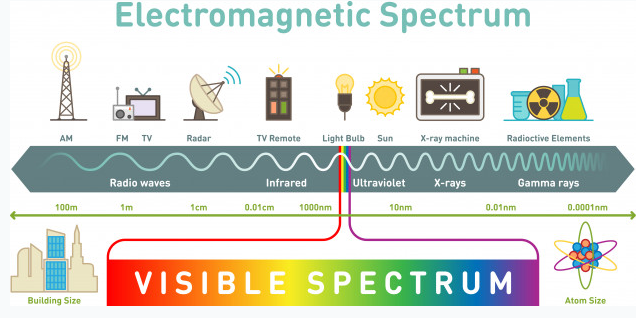
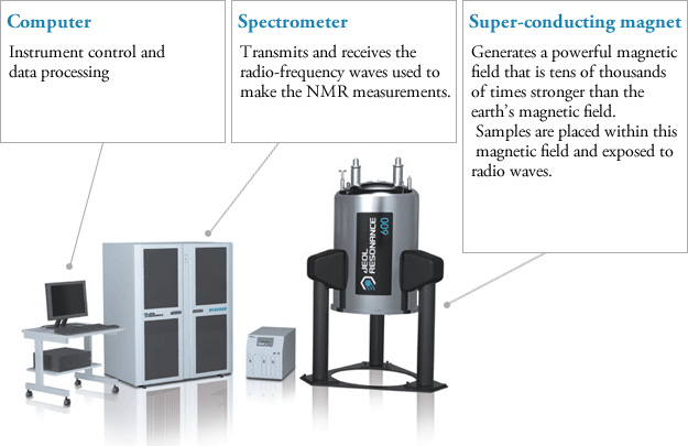
NMR spectroscopy is an illumination method based on the absorption of electromagnetic rays in the radio frequency field by atomic nuclei in a molecule placed in a strong magnetic field. NMR spectroscopy is a technique used in understanding the structure of molecules in the field of chemistry. Hydrogen-containing groups in the molecule and neighbouring groups can also be detected using this method. If evaluated together with the results obtained by other spectroscopic techniques, the structure to be illuminated can be easily reached. In UV and IR spectroscopy, the molecule’s functional groups and the percentages of C, H, O, N, and S atoms in the elemental analysis are determined. NMR spectroscopy gives information about the skeleton of the molecule. Other spectroscopic methods deal with electrons, but NMR spectroscopy deals with the nucleus. NMR requires a strong magnetic field and radio waves, which are long-wavelength rays of the electromagnetic spectrum. NMR spectroscopy does not disrupt the molecule, and analysis samples can be used repeatedly as in UV and IR spectroscopy.

Every atom whose atomic number or mass number is odd has a nuclear spin. The nucleus rotates around itself and is electrically charged creating its magnetic field. Rotating protons behave like bar magnets when placed in an external magnetic field. Rotating protons’ own magnetic fields go either in the same direction with the outer field or in the opposite direction. With the absorption of a photon with a certain amount of energy, the direction of the proton field can change. The energy difference between the two states is directly proportional to the strength of the magnetic field. Protons are surrounded by electrons that protect them from the external magnetic field. Rotating electrons create an exciting magnetic field opposite the external magnetic field and reduce the external field’s influence.

Charged particles rotating around their own axis create electrical and magnetic fields. When these atoms are placed in a stronger magnetic field, two magnetic spins called +1/2 and -1/2 occur. Depending on the direction of the outer magnetic field, the magnetic field due to the atom’s own spin is either added or removed. Thus, the higher and lower energy nucleus can be found.

The small magnetic field in the core and the large applied magnetic field cause the difference in the results of subtraction and addition to be small. This difference changes with the applied external magnetic fields and becomes zero if the magnetic field is not applied. NMR spectroscopy is based on trying to equalise the difference from the external magnetic field with radio frequencies. 

Magnetic Properties of the Nucleus and the Basis of NMR Spectroscopy

Some atomic nuclei act like magnets rotating around themselves forming the basis of NMR spectroscopy. Atomic nuclei are positively (+) charged. The nucleus rotates around itself, and the (+) load moves in orbit around the axis, this is the spin motion. Because the nucleus rotates around itself, it also has angular momentum. A dipole and a magnetic field arise from the spin motion. The size of the dipole is called the nuclear magnetic moment (μ), and the angular momentum of the charge is called the spin quantum number (I). To study the NMR of an element, the magnetic moment must be non-zero (μ≠0), and the spin quantum number must be greater than zero (I>0). The spin quantum number varies according to the number of protons and neutrons in the nucleus. The spin number can be 0, 1/2, 1, 3/2, 5/2. If I=0, there is no spin. Protons and neutrons have their own spins, and their sum gives the number of spins in the nucleus. Isotopes of an element have different spin quantum numbers.

Proton NMR Spectroscopy

NMR spectroscopy, like other spectrophotometers, examines samples in dilute media. First, the sample solution to be NMR is taken into a glass tube of 5 mm in diameter and 15 cm in size and placed in a strong magnetic field. Then the radio frequency is sent to the sample. After the sample emits the radio frequency it has absorbed, the detector measures the re-emitted frequency. This change is related to the externally applied magnetic field, and the applied magnetic field must be kept constant throughout the measurement.

nmr
Components of NMR instrument (Alqaheem Y, Alomair AA: Microscopy and Spectroscopy Techniques for Characterization of Polymeric Membranes. Membranes (Basel) 2020; 10.)
NMR instrument composition

Protons behave differently from each other, which is explained by the electron density around them. Although the proton’s magnetic properties are equal, it is accepted that they will differ in the magnetic environment due to the electron density around it, thus making it easier to understand the spectrum that NMR spectroscopy will give. This difference is minimal compared to the magnetic field and is expressed using ppm (parts per million). Generally, the frequency scale is used instead of magnetic field differences in NMR. The proton resonances of organic molecules are between 0-12 ppm. Highly sensitive devices and systems are required to examine this small range.

The Determination of the Structure of Membrane Proteins using NMR Spectroscopy

Membrane proteins are a part of the biological membranes. They are branched into several types. They can penetrate the cell or be on its surface and have a temporal interaction. Membrane proteins have an important role in the medical area. They are the target structures of the drug. Besides, they have a crucial impact on human and animal diseases. Membrane proteins have the capability of mutating and truncating. While these procedures, several diseases can occur in both humans and animals. 

Membrane proteins have a wide range of characteristics, such as being a receptor and interacting with hormones. They can reveal as enzymes and bind to the receptors. Some of them are membrane transporter proteins and impact the transportation of small molecules, macromolecules, and some ions. Their secondary structure is composed mainly of β-barrel, a β-sheet consisting of a first and last strand bonded with hydrogen bonds.

nmr
18-strand β barrel.

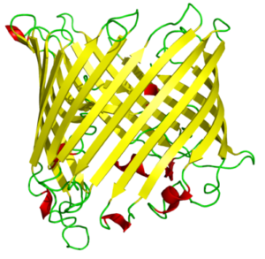
β-barrels mostly can dissolve in water. Strands of the beta barrels contain polar and nonpolar amino acids which lead the protein to have hydrophobic and hydrophilic sides; mostly the hydrophilic part interacts with the solvent because it is a settled surface. The hydrophobic part plays inside of the protein molecule. In proteins containing beta barrels, the hydrophobic side is towards the exterior. They interact with the lipids under the vicinity of it, which surround the protein’s exterior.

liposome, micelle, bilayer sheet
The structures formed by phospholipids in aqueous solutions. Micelles are single-chain lipids.

GPCRs (G-protein coupled receptors), membrane proteins, are cell signal transportation receptors. The 3D structure of these proteins is determined by NMR spectroscopy. In a region made out of lipids, we can observe how membrane proteins’ motions are reflected. It can be observed in a small area or a large area. As the scientists are supposed to dissolve the substance to mark it on the NMR spectroscopy, they use detergents to dissolve, to denature proteins. 

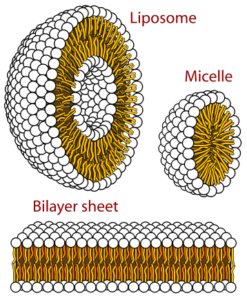
While solubilizing the proteins using detergents, some substances occur—for example, micelles and isotropic bicelles. Micelles are the strewn surface-active molecules’ flocculation. Isotropic bicelles are for the reconstruction of membrane proteins. They patch the proteins with lipid bilayers. Detergents are unnatural substances, so they cause the risk of subverting the protein structure. You can most clearly study the proteins when they are in a phospholipid environment.

 The helical membrane proteins can be observed at a high resolution under the bilayers’ favour. They are in an active and immobilised state. On the NMR, substances are well determined in a high-resolution solid state. Bacteriorhodopsin is the first determined helical membrane protein under NMR spectroscopy. It was solubilised with octyl glucoside, which is a type of detergent. Other than NMR spectroscopy, X-rays have also contributed to the determination of membrane proteins. This type of protein was first observed under x-ray diffraction. It was observed during a photosynthetic reaction. The product of this reaction was Rhodopseudomonas viridis (a kind of bacteria), and it was solubilized in a powerful detergent, N, N-dimethyl dodecyl amine N-oxide. The detergent is composed of amphiphilic (both have the characteristic of being hydrophobic and hydrophilic) compounds.

Carbohydrate-Protein Interactions

NMR can identify carbohydrate-protein interactions with the resolution in solvents. This interaction is necessary for the virus-to-cell and cell-to-cell connection. Continuous interaction is provided by the carbohydrate-protein mutual effect for an infection or adhesion event. Even in viruses and pathogens, the outside of the cell is surrounded by glycans. Recognisance and biological transactions are instructed by the mutual effect of the glycans and protein receptors. A variety of molecules can have a mutual effect on carbohydrates. It happens in the vicinity of a mutual effect between L-selectins and glycans terminated by sialic acid.

Influenza infection requires binding to carbohydrates. Hemagglutinin cells (influenza virus) have to be connected to the Siaα-6Gal. The adhesion and growth of tumour cells are supported by β-lactosamine’s mutual effect, which includes galectins and glycans. There are several approaches to NMR. It can be protein-based or ligand-based. A ligand is a complex compound that contains attached biomolecules. If it is protein-based, it will have different solubility characteristics from other solutions. High-resolution attainability requires labelling the protein with C and N stable isotopes. By this labelling procedure, the size will be in the range of 10-25 kDa, which is in NMR standards. There are some protein-detecting methods. In the protein’s 3D structure, the scientist must map the linkage regions of the carbohydrate onto it. The linkage regions are determined by the correlation spectra of H and N atoms. Ligands of carbohydrates must be defined because they are specified from various proteins. In terms of the NMR method (methods of protein detection), it is crucial to compare the proteins in their free state and bound to the carbohydrate state. The labelled protein quantity is dependent on several topics. The first one is the instrumentation of NMR, and the second one is the spectrometer’s available time.

NMR Spectroscopy for the Assignation of Unsaturated Fatty Acids

Fatty acids (FAs) are usually identified by gas chromatography (GC). FAs have to be transformed into methyl esters to assign them to GC. There are saturated and unsaturated FAs. Unsaturated FAs (UFAs) carry one, two or more double bonds and are critical components of animal fats and vegetable oils. UFAs having two or more double bonds are referred to as Polyunsatured FAs (PUFAs). There is a method based on the carbon NMR To determine these bonds’ percentages. Besides, scientists also use hydrogen to quantify UFAs.

Chemical Shift

NMR signals change depending on the magnetic field and radio frequencies. Therefore, radiofrequency change is studied in a standard magnetic field or in a magnetic field change under a standard radio frequency. This problem is avoided by adding a standard substance during NMR measurements. This substance should not affect the hydrogen atom’s electron density with its electronegativity much, and it is tetramethylsilane (TMS) without a solubility problem. All the hydrogens of TMS are equal and at the same point. This point is referenced (point O). The interpretation of where other hydrogen atoms come out according to this point is called the chemical shift. Its unit is ppm, and the  symbol represents it. During NMR measurements, it is preferred that the molecules whose spectrum is to be taken be diluted. The presence of hydrogen atoms in the solvent causes the problem of intensity. Some solvents do not contain hydrogen, and generally, not all substances have good solubility. To solve this problem, deutero structures that are not affected by the magnetic field are used. The most common and first tried solvent is deutero chloroform. Solvents such as deutero water, ethyl alcohol, and dimethylsulfoxide are also widely used.

Proton chemical shift values

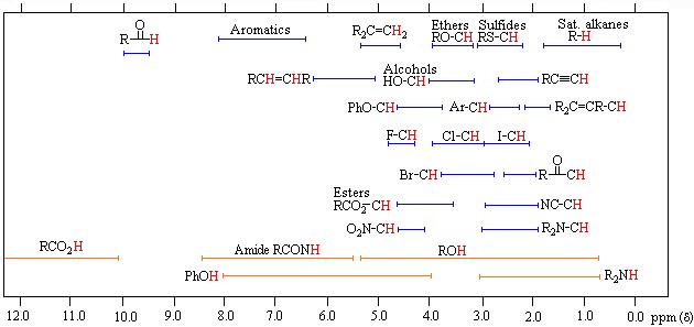
Before making the NMR spectrum interpretation, the electron density around the hydrogen atom must be known from a rough perspective. Compared with TMS, the electron density around a hydrogen atom changes depending on the electronegativity and shielding effect of the atoms to which it is attached. If we accept TMS=0 ppm, Si electronegativity is less than C electronegativity, so the electron density in hydrogen atoms bound to carbon atoms will be less than TMS and shielding less. Thus, we see that radio frequency signals reach the protons in the nucleus and return; thus, excitation and resonance will be easier. It requires less magnetic field, so the ppm gradually increases from 0. Therefore, C-H’s are attached to the carbon atom in molecules that do not contain electronegative, such as 0-1. The chemical shift of hydrogen atoms with low electron density and shielding around them, such as the RCOOH acid proton, is as large as 11–12 ppm. Although there are tables for proton chemical shift values, it is often possible to approximate peaks and locations. It is not always necessary to look at this crowded picture, but it may be necessary with some complex and large molecules.

In the NMR spectrum, the signal intensity depends on the substance concentration. Dilute solutions give weak signals. If the concentration increases, the peak intensity increases. If we take the peaks of different substances at the same concentration, we see that the peak intensity depends on the equivalent hydrogen number. If we take the equivalent concentration of benzene containing 6 equivalents of hydrogen and cyclohexane containing 12 equivalents of hydrogen and measure the NMR spectrum, the NMR spectrum peak intensity of the cyclohexane is twice that of benzene. When these results are combined with the knowledge of how many hydrogen atoms it contains and chemical shift, it makes it easier to understand where the hydrogens of the molecule come out. However, there are still some problems with knowing how many hydrogens each peak equals. However, hydrogen numbers can be calculated from the ratio of peaks to peaks, but instead of exact numbers, they are found in coefficients relative to each other. It is possible to estimate approximate values with this integration method, similar to a simple molecular formula calculation.

In conclusion, NMR spectroscopy has contributed to the determination of various complex molecules like proteins, carbohydrates, enzymes, intracellular metabolites, receptors, and transporters. Understanding these structures by scientists leads to the development of science. Complex molecules are essential to understand various diseases, both in humans and animals. Although NMR is complicated for us, it is a beneficial thing today. That’s why we should try to grasp a complex subject’s logic rather than make it difficult in our minds. The blessings of NMR are countless. It is used in many industries, such as polymer research, synthetic chemistry, petrochemistry, biochemistry, textiles, food, paint, medicine, and agriculture. We can achieve many things using NMR, such as the compound’s nature, structure shape and bonding structure, mixture components, atomic composition, molecular weight and formula, polymer composition and arrangement, and molecular motion. No matter how boring its theory may sound, we do not know how correct it is to call something ‘boring’ that we use somehow.


This article is a part of the home assignment written by Ada Begüm Ögel and Emir Kerem Demiroğlu, two of our 2020-2021 Academic Year Fall Semester Organic Chemistry students.


References

  • Alqaheem Y, Alomair AA: Microscopy and Spectroscopy Techniques for Characterization of Polymeric Membranes. Membranes (Basel) 2020; 10.
  • Aletli Analiz Yöntemleri: Nükleer Manyetik Rezonans (NMR) Spektroskopisi [http://web.hitit.edu.tr/dersnotlari/gokcemerey_13.10.2015_7A4L.pdf]
  • Granger RM, Yochum HM, Granger JN, Sienerth KD: Instrumental Analysis: Revised Edition. Oxford University Press; Revised edition; 2017.
  • Nükleer Magnetik Resonans Spektroskopisi (NMR) [http://w3.balikesir.edu.tr/~hnamli/oya/nmr/hnmr.php]
  • Staudacher T, Shi F, Pezzagna S, et al.: Nuclear magnetic resonance spectroscopy on a (5-nanometer)3 sample volume. Science (80- ) 2013; 339:561–563.
  • Siegal G, Selenko P: Cells, drugs and NMR. J Magn Reson 2019; 306:202–212.
  • Shulman GI, Alger JR, Prichard JW, Shulman RG: Nuclear magnetic resonance spectroscopy in diagnostic and investigative medicine. J Clin Invest 1984; 74:1127–1131.
  • Bewley CA, Shahzad-ul-Hussan S: Characterizing carbohydrate-protein interactions by nuclear magnetic resonance spectroscopy. Biopolymers 2013; 99:796–806.
  • Wider G: Structure determination of biological macromolecules in solution using nuclear magnetic resonance spectroscopy. Biotechniques 2000; 29:1278–82, 1284–90, 1292 passim.
  • Campagne S, Gervais V, Milon A: Nuclear magnetic resonance analysis of protein-DNA interactions. J R Soc Interface 2011; 8:1065–1078.
  • Simpson MJ, Hatcher PG: Determination of black carbon in natural organic matter by chemical oxidation and solid-state 13C nuclear magnetic resonance spectroscopy. Org Geochem 2004; 35:923–935.
  • Opella SJ, Marassi FM: Structure determination of membrane proteins by NMR spectroscopy. Chem Rev 2004; 104:3587–3606.
  • Miyake Y, Yokomizo K, Matsuzaki N: Determination of unsaturated fatty acid composition by high-resolution nuclear magnetic resonance spectroscopy. J Am Oil Chem Soc 1998; 75:1091–1094.
  • Chalbot M-CG, Kavouras IG: Nuclear magnetic resonance spectroscopy for determining the functional content of organic aerosols: a review. Environ Pollut 2014; 191:232–249.
  • Zia K, Siddiqui T, Ali S, Farooq I, Zafar MS, Khurshid Z: Nuclear Magnetic Resonance Spectroscopy for Medical and Dental Applications: A Comprehensive Review. Eur J Dent 2019; 13:124–128.

Potential lymphoma marker in dogs and cats: Thymidine kinase activity

The incidence of non-Hodgkins Lymphoma (NHL) or Malignant Lymphoma (ML) in dogs is reported to be more than 24 per 100,000. Advances in the diagnosis and treatment of ML in dogs not only improve the quality of life of animals but also enable better models in veterinary comparative oncology.

Thymidine kinase (TK) is an intracellular enzyme that plays an important role during pyrimidine synthesis. TK activity increases markedly in the G1-S phase, especially during cell division, and decreases rapidly in the G2 phase. Therefore, high extracellular TK activity reflects high DNA synthesis and cells that die during cell division. Hematopoietic system malignancies are characterized by high cell proliferation. Studies in the veterinary field have shown that serum Thymidine kinase activity is an important marker in the diagnosis, prognosis and monitoring of treatment efficacy in leukaemia, multiple myeloma and malignant lymphoma.

TK activity has been used for years in the diagnosis, prognosis and treatment follow-up of hematopoietic tumours in human oncology, and the first study in the veterinary field was conducted by Nakamura et al. conducted in 1997 in Lymphoma, leukaemia, non-hematopoietic tumours (breast tumour, mastocytoma, anal sac tumour, malignant histiocytosis) and healthy dogs. After the analysis, TC activity in dogs with Lymphoma and Leukemia was significantly increased compared to healthy dogs; in dogs with non-hematopoietic tumours, it was found to be at the same level as healthy dogs. Again in the same study, it was determined that TC Activity is important in the follow-up of the treatment in the analyzes performed before the treatment, at the stage of the disappearance of clinical symptoms, and at the relapse stage.

timidin kinaz, köpek, lenfoma, Thymidine kinase

In another study conducted by Prof. Dr. Hendrik von EULER et al. in dogs diagnosed with ML between 1999-2003, it was reported that TK Activity can be used as a strong marker in the diagnosis of ML disease, especially in determining the prognosis and predicting clinical disease before a recurrence in dogs undergoing chemotherapy. Serum TK Activity was found to be 2 to 180 times higher in dogs with ML disease than in healthy dogs. It was determined that TC activity decreased to normal values ​​in dogs that responded to treatment and whose cancer symptoms disappeared (complete remission), and TC activity increased again before recurrence. In the same study, it was determined that TC activity was correlated with the clinical stages of the disease.

Similar studies have been carried out in cats in recent years, along with studies in dogs. The first study on cats was conducted on a total of 171 cats in the UK and Sweden, published in 2012 and also included in our partner laboratory, Dechra Specialist Laboratories. Of the cats included in the study, 49 were healthy, 33 had lymphoma, 55 had the inflammatory disease, and 34 had non-hematopoietic neoplasia. At the end of the study, it was determined that the serum TC activity was significantly higher in cats with lymphoma compared to the others, and it was reported that high TC activity would strengthen the diagnosis of lymphoma.

Thymidine kinase activity with recent studies;

  • In the diagnosis of lymphoma and leukaemia together with other clinical and laboratory findings,
  • In evaluating the prognosis,
  • In the evaluation of chemotherapeutic success with analyzes performed before, during and after treatment,
  • Monitoring chemotherapy and identifying relapse cases before they form,
  • It has been used successfully in distinguishing clinical worsening in patients receiving chemotherapy treatment.

References

  • Boyé, P. et al. (2019) ‘Evaluation of serum thymidine kinase 1 activity as a biomarker for treatment  effectiveness and prediction of relapse in dogs with non-Hodgkin lymphoma.’, Journal of veterinary internal medicine, 33(4), pp. 1728–1739. doi: https://doi.org/10.1111/jvim.15513.
  • Bryan, J. N. (2016) ‘The Current State of Clinical Application of Serum Biomarkers for Canine Lymphoma.’, Frontiers in veterinary science, 3, p. 87. doi: https://doi.org/10.3389/fvets.2016.00087.
  • von Euler, H. et al. (2004) ‘Serum thymidine kinase activity in dogs with malignant lymphoma: a potent marker for  prognosis and monitoring the disease.’, Journal of veterinary internal medicine. United States, 18(5), pp. 696–702. doi: https://doi.org/10.1371/journal.pone.0137871.
  • Kayar, A. et al. (2018) ‘Clinical features, haematologic parameters, blood serum biochemistry results and thymidine kinase activity of dogs affected by malignant lymphoma in Turkey’, Japanese Journal of Veterinary Research, 66(4), pp. 227–238. doi: https://doi.org/10.14943/jjvr.66.4.227.
  • Larsdotter, S., Nostell, K. and von Euler, H. (2015) ‘Serum thymidine kinase activity in clinically healthy and diseased horses: a  potential marker for lymphoma.’, Veterinary journal (London, England : 1997). England, 205(2), pp. 313–316. doi: https://doi.org/10.1016/j.tvjl.2015.01.019.
  • Nakamura, N. et al. (1997) ‘Plasma thymidine kinase activity in dogs with lymphoma and leukemia.’, The Journal of veterinary medical science. Japan, 59(10), pp. 957–960. doi: https://doi.org/10.1292/jvms.59.957.
  • Selting, K. A. et al. (2016) ‘Thymidine Kinase Type 1 and C-Reactive Protein Concentrations in Dogs with  Spontaneously Occurring Cancer.’, Journal of veterinary internal medicine, 30(4), pp. 1159–1166. doi: https://doi.org/10.1111/jvim.13954.
  • Taylor, S. S. et al. (2013) ‘Serum thymidine kinase activity in clinically healthy and diseased cats: a potential  biomarker for lymphoma.’, Journal of feline medicine and surgery. England, 15(2), pp. 142–147. doi: https://doi.org/10.1177/1098612X12463928.

The timeless method: The Jaffe Reaction

Jaffe_Reaction

Blood creatinine concentration is measured for purposes such as diagnosis of kidney failure, determination of its stage, follow-up of treatment, and evaluation of prognosis.

In addition, from time to time, urine creatinine concentration is measured and evaluated alone or in combination with other test parameters (such as protein). The only way to make these measurements is to use specific analytical methods. One of the commonly used methods is the Jaffe reaction. The creatinine concentration is determined from blood and urine samples by the Jaffe reaction, which is a colorimetric method1.

Max Jaffe

132 years ago (1886) Max Jaffe (1841-1911) discovered that creatinine reacts with picric acid in an alkaline environment and explained this by publishing his article “Über den Niederschlag, welchen Pikrinsäure in normalem Harn erzeugt und über eine neue Reaction des Kreatinins”2. The article describes this reaction and the nature of the precipitate formed. Jaffe’s discovery was a turning point. As a result of this study, the method of measuring creatinine concentration, which has become extremely popular and defied time, was born.

Over time, Jaffe’s name became synonymous with clinical creatinine testing, although his article later became the permanent method and the principle of further studies. At the beginning of the twentieth century, Otto Folin (1867-1934), taking up the research of Max Jaffe, developed a colorimetric method for measuring the concentration of creatinine in blood and urine3 and made it into modern biochemistry analysis.

Although there are more specific analytical methods4 today, this unique test is still used as the preferred method due to its simplicity of implementation, speed, compatibility with automated analyzers, and cost-effectiveness. Besides, the Jaffe reaction is the oldest test method used in clinical laboratories.


References

  1. Delanghe JR, Speeckaert MM. Creatinine determination according to Jaffe – What does it stand for? NDT Plus. 2011;4(2):83-86. doi: http://doi.org/10.1093/ndtplus/sfq211
  2. Jaffe M. Ueber den Niederschlag, welchen Pikrinsäure in normalem Harn erzeugt und über eine neue Reaction des Kreatinins. ZPhysiolChem. 1886. doi: https://doi.org/10.1515/BCHM1.1886.10.5.391
  3. Folin O. Beitrag zur Chemie des Kreatinins und Kreatins im Harne. Hoppe Seylers Z Physiol Chem. 1904. doi: https://doi.org/10.1515/bchm2.1904.41.3.223
  4. Panteghini M, IFCC. Enzymatic assays for creatinine: Time for action. Scand J Clin Lab Invest. 2008;46(4):567-572. doi: https://doi.org/10.1080/00365510802149978

Total calcium (tCa), Ionized calcium (iCa), Corrected total calcium (ctCa): Which?

Calcium (Ca) is one of the macro elements that have great importance in both animal and human metabolism.

In the regulation of total calcium (tCa) metabolism, mainly skin, liver, kidneys, bones and intestines at the tissue level; Parathyroid Hormone (PTH), Calcitonin (CT) and vitamin D take part at the molecular level. Calcium is the structural component of the skeletal system and has different and various functions in the organism. These include muscle contraction, blood coagulation, enzyme activity, neural stimulation, hormone release, secondary messenger, and membrane permeability.

The calcium ion concentration of the extracellular fluid in the body is vital and is always kept in balance. Parathyroid hormone (PTH), Calcitonin (CT) and Vitamin D contribute primarily to this balance. Apart from these, other hormones such as adrenal corticosteroids, estrogens, thyroxine, somatotropin and glucagon also contribute.

Calcium in plasma or serum is divided into 3 fractions. These:

  1. Ionized or free calcium (iCa or Ca++) (≈56%)
  2. Protein-bound calcium (mostly albumin) (≈34%)
  3. Complex or chelated calcium (transports bound to various anions with small molecular weights-phosphate, bicarbonate, citrate, lactate) (≈10%)

iCa and complexed calcium form the dispersible fraction of calcium. This fraction may also be referred to as ultrafiltrate calcium as it passes through biological membranes. iCa is the most physiologically active fraction of serum calcium. iCa is responsible for functions such as bone homeostasis, nerve conduction, blood coagulation, Vitamin D and PTH secretion, activation of metabolic and digestive enzymes, and effective use of iron, and is also a sensitive marker of pathological conditions.

About 90% of protein-bound calcium is bound to albumin and the remaining 10% is bound to various globulins. Since approximately half of the calcium is bound to proteins, the evaluation of tCa depends on serum albumin and total protein values (Figure 1).

calcium, ionized calcium

Figure 1. While iCa normally remains in a very narrow range, the tCa concentration is affected by either bound or complex calcium. In other words, the tCa concentration may differ depending on the change in protein-bound Ca or complex Ca fractions.

Traditionally, the assessment of an animal’s calcium status has been based on the assessment of its tCa concentration. The tCa concentration is assumed to be directly proportional to the biologically active fraction and iCa, the gold standard for the determination of calcium status. However, this assumption is not valid in a variety of clinical situations. It has been suggested that tCa can be corrected or adjusted according to albumin or total protein concentration to improve the diagnostic interpretation, especially in patients with hypoalbuminemia or hypoproteinemia, when iCa measurement is not possible. Also, changes in pH change the calcium fraction bound to albumin; therefore, the iCa concentration can also change without a change in tCa. This corrected or adjustable tCa is called corrected calcium (ctCa). Evaluation of ctCa is recommended, especially when the plasma albumin concentration changes.

Concentration measurement of tCa, albumin and total protein can be done easily with in-house and laboratory-type analytical devices. Generally, Arsenazo III, Bromcresol green and Biuret methods are used in this type of device, respectively.

The measurement of iCa concentration is made with devices with ion-selective electrodes (ISE). Such devices can be mobile or bench-type POC (point-of-care) devices (Figure 2), or they can be a component of automated biochemistry analyzers. Mobile-type devices are frequently preferred in clinics and are often costly, and it is recommended to compare the results with reference laboratory results; This process is recommended in doubtful cases or for quality control purposes from time to time.

When measuring iCa concentration, sample collection and processing should be done with the utmost care and attention. Samples should be collected in an anaerobic environment (to minimize carbon dioxide loss), transported in the cold chain and processed within a few hours (to minimize lactate production). tCa concentration measurements are relatively inexpensive, readily available, and more robust to sample transport variables. For these reasons, the measurement of total calcium is frequently performed and evaluated today.

As a result, it is reported that all three parameters can be used in monitoring the body’s Ca balance. The most important thing at this point is to understand what each parameter is, its variables and what could be misleading. The use of tCa as an indicator of Ca status, especially in hypoalbuminemia cases, tends to overestimate hypocalcemia and ignore normocalcemia; Using ctCa may result in overestimating normocalcemia and ignoring hypocalcemia. Therefore, it is recommended to evaluate Ca homeostasis with iCa concentrations instead of tCa or ctCa in hypoalbuminemia cases. Thus, it can be determined whether there is true hypocalcemia.


References

1-Caprita R, Caprita A, Cretescu I. Estimation of Ionized Calcium and Corrected Total Calcium Concentration Based on Serum Albumin Level. Anim Sci Biotechnol. 2013;46(1):180-184.
2-Danner J, Ridgway MD, Rubin SI, Le Boedec K. Development of a Multivariate Predictive Model to Estimate Ionized Calcium Concentration from Serum Biochemical Profile Results in Dogs. J Vet Intern Med. 2017;31(5):1392-1402. doi: https://doi.org/10.1111/jvim.14800
3-Bohn AA. Veterinary Hematology and Clinical Biochemistry. 2nd ed. (Thrall MA, Weiser G, Allison R, Campbell T, eds.). NJ, US: Wiley-Blackwell, John Wiley & Sons; 2012.
4-Payne RB, Carver ME, Morgan DB. Interpretation of serum total calcium: effects of adjustment of albumin concentration on frequency of abnormal values and on detection of change in the individual. J Clin Pathol. 1979;32(1):56-60. doi: https//doi.org/10.1136/jcp.32.1.56
5-Sharp CR, Kerl ME, Mann FA. A comparison of total calcium, corrected calcium, and ionized calcium concentrations as indicators of calcium homeostasis among hypoalbuminemic dogs requiring intensive care: Original study. J Vet Emerg Crit Care. 2009;19(6):571-578. doi: https//doi.org/10.1111/j.1476-4431.2009.00485.x
6-Toffaletti JG. September 2011 Clinical Laboratory News : Calcium. 2011;37(9):6-10.

Bisphenol A (BPA) Exposure: Potential Hazard in Canned Pet Foods

bisphenol-A

Bisphenol A (BPA; IUPAC name: 2,2-bis(4-hydroxyphenyl) propane) is an organic synthetic compound that has a dysfunctional effect on the endocrine system.

Bisphenol A (BPA) causes random and systemic effects in living things without tissue separation. They can block the binding of natural ligands to their respective receptors. For example;

  • Changes in the activity of gonadal hormones.
  • Disturbances in thyroid hormone function; It is structurally similar to thyroid hormones and acts as a thyroid hormone receptor antagonist.
  • Differences in central nervous system function.
  • Suppression of the immune system.
konserve mama, bisfenol a, bisphenol a

BPA is a chemical used in the manufacture of many household items. It is frequently used in food and beverage packaging materials; polycarbonate plastics (plastic bottles, storage containers…), and epoxy resins (metal cans, soft drink cans, aluminum containers, tin containers…). Today, the production volume of BPA in the world is expressed in trillions of dollars annually and this volume is increasing every year.

Due to the faulty production of containers containing BPA, BPA is migrated to the food and it is taken into the body with the consumption of this food. Considering its negative impact on living things and its widespread use, its importance emerges and it draws attention, especially in packaging products that come into contact with canned food. Due to its negative effects on human health, many countries, especially the United States, the European Union, and the Republic of Turkey (Turkish Food Codex Communiqué on Plastic Substances and Materials in Contact with Food; No: 2013/34), have limited the use of BPA and set migration limits. In addition, its use is completely prohibited, especially in some products; such as polycarbonate baby bottles, pacifiers, and bottle caps. It is seen that these legal regulations and many studies are completely focused on human health.

Bisphenol A and Pets

It is known that cats and dogs are mostly fed commercial foods. However, the BPA content of these foods is questionable. Regarding BPA exposure, studies on the packaging of pet foods (especially cans) and food migration, the level of BPA exposure in pet animals, and potential health consequences are limited.

In a study, different concentrations of BPA were detected in 15 different commercial cat foods and 11 different commercial dog foods (Kang and Kondo, 2002).

In a study comparing cats with hyperthyroidism and cats with normal thyroid function, it was determined that feeding canned food poses a higher risk for hyperthyroidism than feeding other types of packaged food. It has been hypothesized that this situation may be related to the BPA content of canned foods (especially aluminum-composition tin containers) (Edinboro et al., 2004; Köhler et al., 2016).

In a recent study on dogs (Koestel et al. 2017), the BPA content in commercial dog foods was determined and the effects on the exposure level and health status of animals were investigated after consumption of these foods in a short time period (two weeks). For this purpose, a two-week feeding program was applied, one using packaged commercial food specified as BPA-free and the other commercial food without such an indication. The serum BPA concentrations of the animals were compared with the hematological tests, serum biochemistry, cortisol, DNA methylation, and intestinal microbiome changes in the samples (blood, feces) taken before the diet and two weeks later. As a result of the study, it was determined that serum BPA concentrations increased 3 times in dogs fed with both foods. It was determined that this increase was accompanied by changes in serum biochemistry and microbiome. It has been determined that the increase in serum BPA level decreases the bacterial species in the microbiome. One of the interesting findings of the study was the detection of measurable levels of BPA, even in foods specified as BPA-free.

BPA can bioaccumulate in terrestrial and aquatic resources, thus posing the risk of continued exposure to animals and humans. In a world where humans and animals live together, we cannot separate human and animal health. The principle of “One Health” should always be adopted, the issue of BPA should be approached from this perspective and more studies on the subject are needed.


References

  • Edinboro CH, Scott-Moncrieff JC, Janovitz E, Thacker HL, Glickman LT, (2004). Epidemiologic study of relationships between consumption of commercial canned food and risk of hyperthyroidism in cats. J Am Vet Med Assoc., 224(6):879-886. https://doi.org/10.2460/javma.2004.224.879
  • Kang JH, Kondo F, (2002). Determination of bisphenol A in canned pet foods. Res Vet Sci., 73(2):177-182. https://doi.org/10.1016/s0034-5288(02)00102-9
  • Koestel ZL, Backus RC, Tsuruta K, Spollen WG, Johnson SA, Javurek AB, Ellersieck MR, Wiedmeyer CE, Kannan K, Xue J, Bivens NJ, Givan SA, Rosenfeld CS, (2017). Bisphenol A (BPA) in the serum of pet dogs following short-term consumption of canned dog food and potential health consequences of exposure to BPA. Sci Total Environ., 579:1804-1814. https://doi.org/10.1016/j.scitotenv.2016.11.162
  • Köhler I, Ballhausen BD, Stockhaus C, Hartmann K, Wehner A, (2016). Prevalence of and risk factors for feline hyperthyroidism among a clinic population in Southern Germany. Tierarztl Prax Ausg K Kleintiere Heimtiere., 44(3):149-157. https://doi.org/10.15654/tpk-150590
  • Er B, SarımehmetoğluB, (2011). Gıdalarda bisfenol A varlığının değerlendirilmesi. Vet. Hekim Der Derg., 82(1):69-74.